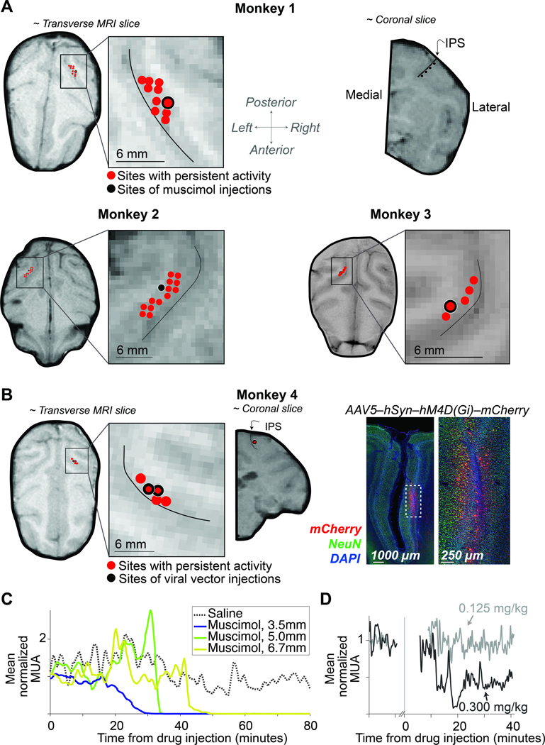
Figure 2: Localization and characterization of LIP inactivation sites.
A, The locations of muscimol injections and neurons with spatially selective persistent activity are superimposed on MRIs for Monkeys 1, 2, and 3. The near-transverse planes are orthogonal to the injection trajectories. The near-coronal MRI slice from Monkey 1 (top-right) shows positions along the intraparietal sulcus (IPS) where muscimol was injected. The thin black curve (inset) marks the center of the IPS. B, Left: Location of viral vector injections for Monkey 4. The red points in the MRI are sites containing neurons with spatially selective persistent activity. The coronal slice shows the injection site; same conventions as in A. Right: Representative histology. Expression of hM4Di-mCherry receptor is restricted to the lateral bank of the IPS. C, Time course of multi-unit activity (MUA) in area LIP following injection of saline (dotted) and muscimol (solid). Recordings were obtained at different distances from the injection site (legend). Note the complete suppression of activity in < 1 hour. The post-drug testing session began 15 minutes after the completion of muscimol infusion (at least 1 hour after the start of the infusion). See also Figure S2. D, Time course of MUA following subcutaneous injection of clozapine at the lowest (gray) and highest (black) dose tested.

