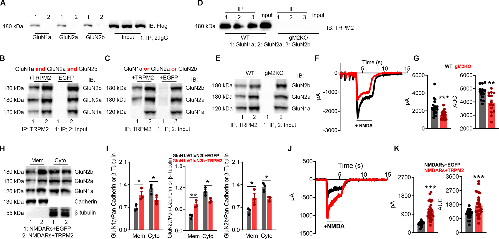
Figure 2 |. TRPM2 physically and functionally interacts with NMDARs.

(A-B), Co-IP of NMDARs and TRPM2 expressed in HEK-293T cells. (A), immunoprecipitation (IP) using anti-NMDARs and IB with anti-Flag. (B), IP using anti-Flag and IB with anti-NMDARs.
(C), Co-IP of TRPM2 co-expressed with GluN1a, GluN2a, or GluN2b in HEK-293T cells. IP using anti-Flag and IB with anti-NMDARs.
(D-E), Co-IP of NMDARs and TRPM2 in the brain lysates. (D) IP using anti-NMDARs and IB with anti-TRPM2. (E) IP using anti-TRPM2 and IB with anti-NMDARs.
(F-G), NMDAR current recording from isolated WT and gM2KO neurons. (G), Average peak current amplitude and area under curve (AUC) (n=20/group).
(H-I), Surface expression of NMDARs in HEK-293T cells co-transfected with NMDARs/TRPM2, or NMDARs/EGFP plasmids. Membrane (Mem) and cytosol (Cyto) protein levels assessed with WB.
(J-K), NMDAR current recording from HEK293T cells transfected with NMDARs/TRPM2, or NMDARs/EGFP. (K), Average peak current amplitude and AUC from NMDARs/EGFP group (n=23) and NMDARs/TRPM2 group (n=27).
(*, p < 0.05, **, p < 0.01, ***, p < 0.001; ANOVA, Bonferroni’s test; mean ± SEM,)
