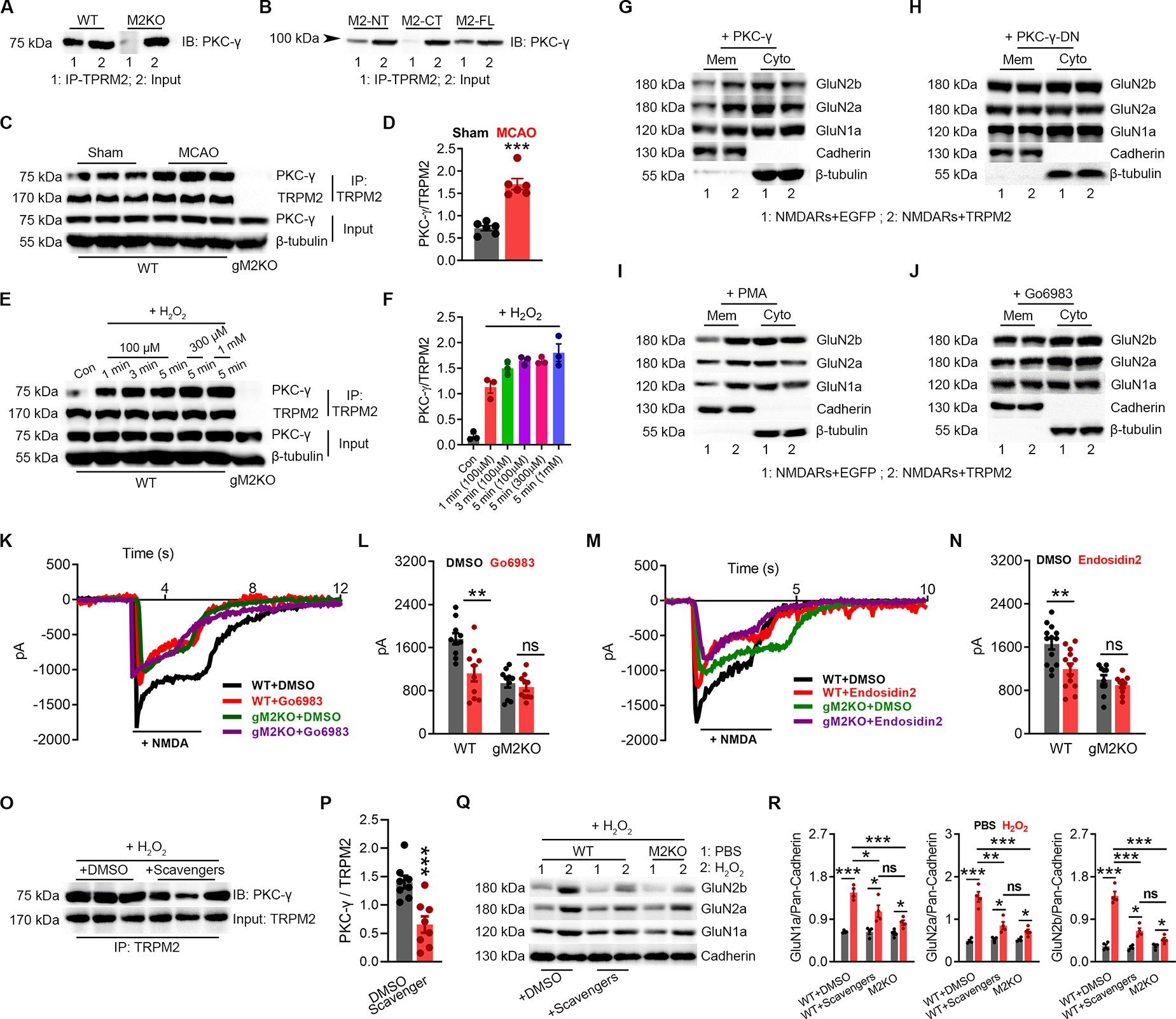
Figure 5 |. N-tail of TRPM2 Interacts with PKC-γ.

(A-B), Co-IP of PKCγ and TRPM2 using brain lysates (A), and in HEK293T cells expressing PKCγ with TRPM2-FL, TRPM2-NT, or TRPM2-CT (B). IP using anti-TRPM2 ( anti-TRPM2-NT for M2-NT, and anti-TRPM2-CT for M2-CT) and IB using anti- PKCγ.
(C-D), Co-IP of TRPM2 and PKCγ using brain lysates from WT mice subjected to MCAO or sham surgery. IP using anti-TRPM2 and IB using anti- PKCγ. (D) Quantification of PKCγ/TRPM2 (n=6/group).
(E-F), Co-IP of TRPM2 and PKCγ using neuron lysates from WT mice subjected to H2O2 treatment (at 100 μM for 1 min, 3 min, and 5 min, at 300 μM for 5 min and at 1 mM for 5 min) (E), IP using anti-TRPM2 and IB using anti-PKCγ. (F), Quantification of PKCγ/TRPM2 (n=3/group).
(G-H), Surface expression of NMDARs in HEK-293T cells co-transfected with PKC-γ/EGFP and PKC-γ/TRPM2 (G) or PKC-γ-DN/EGFP and PKC-γ-DN/TRPM2 (H).
(I-J), Surface expression of NMDARs in HEK-293T cells co-transfected with TRPM2 or EGFP with the treatment of PKC activator PMA (I) or inhibitor Go6893 (J).
(K-L), NMDAR current recording in WT and gM2KO neurons treated with or without Go6983 at 1 μM for overnight. (L), Mean current amplitude (n=10~15 neurons from 2 mice).
(M-N), NMDAR current recording in WT and gM2KO neurons treated with or without the treatment of exocytosis inhibitor endosidin2 at 1 μM overnight. (N) Mean current amplitude (n=10~15 neurons from 2 mice).
(O, P) Co-IP of TRPM2 and PKCγ using neuron lysates from WT mice subjected to H2O2 treatment at 100 μM for 3 min with the preincubation of DMSO or scavengers. (P), Co-IP using anti-TRPM2 and IB using anti- PKCγ. (D) Quantification of PKCγ/TRPM2 (n=6/group).
(Q, R) Surface expression of NMDARs in isolated WT neurons subjected to H2O2 at 100 μM for 3 min with the preincubation of DMSO or scavengers (n=4 in each group).
(ns, no statistical significance, *, p < 0.05, **, p < 0.01, ***, p < 0.001; ANOVA, Bonferroni’s test; mean ± SEM)
