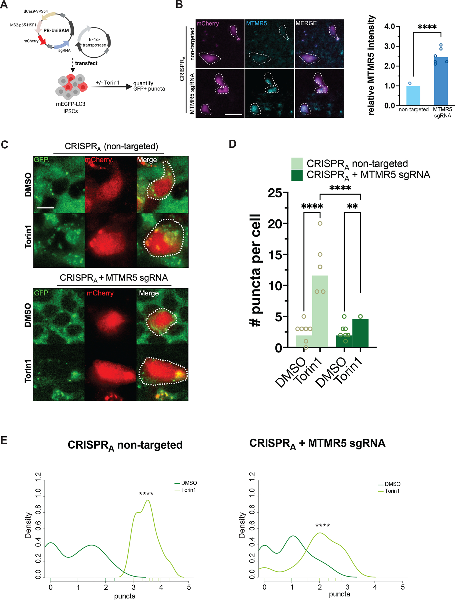
Figure 4. MTMR5 is sufficient to desensitize iPSCs to Torin1 induction of autophagy.

(A) Schematic of CRISPRA experimental workflow using piggybac/transposase system to express dCas9 fused with VPS64-MS2-p65 transactivators. Cells were treated with 250nM Torin1 or vehicle control for 4h then fixed, stained, and imaged. (B) Representative images of iPSCs transfected with CRISPRA vectors without a targeting sgRNA (top) or with sgRNA targeting the native SBF1 locus, followed by immunocytochemistry staining for MTMR5 to confirm protein overexpression. Cells harboring the piggybac/transposase vectors are indicated by the mCherry expression marker. Scale bar, 15μm. ****p<0.0001, Student’s t test. (C) Representative images of mEGFP-LC3-positive vesicles visualized in mCherry-positive cells after treatment with DMSO vehicle or Torin1. Scale bar, 10μm. (D) Scatterplot of blinded manual quantifications of mEGFP-LC3-positive vesicles imaged as in (C). Data are from three independent experiments. **p<0.01; ****p<0.0001, one-way ANOVA. (E) Density plots of data from (D). ****p<0.0001, two-sample Kolmogorov-Smirnov test.
