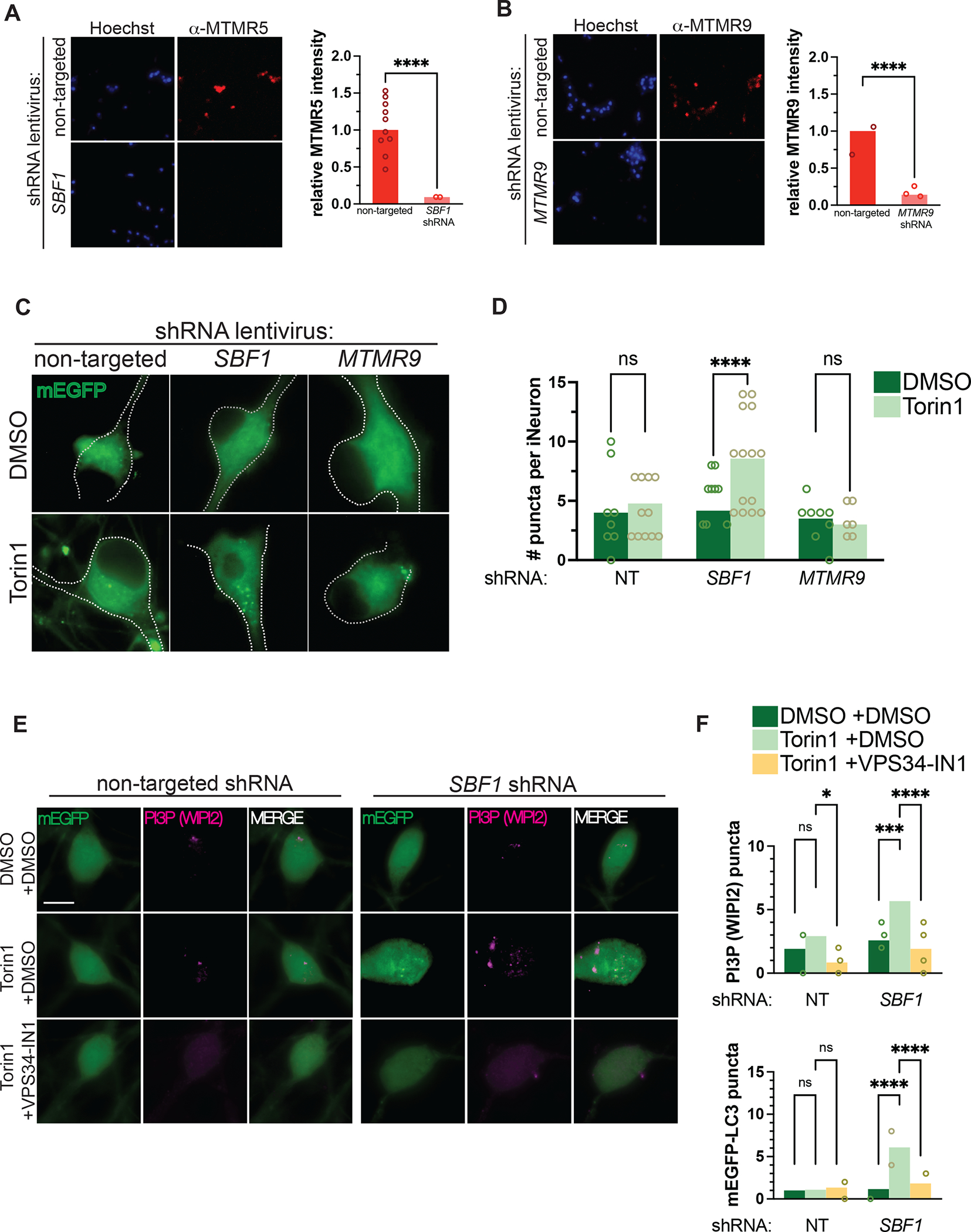
Figure 5. MTMR5 is necessary for suppressing autophagy in neurons.

(A–B) Representative immunocytochemical staining against MTMR5 (A) and MTMR9 (B) in iNeurons transduced with non-targeted shRNA lentivirus (top rows), SBF1 shRNA (bottom left row), or MTMR9 shRNA lentivirus (bottom right row), respectively. (C) Representative images of DIV14 iNeurons transduced with non-targeted, SBF1, or MTMR9 shRNA and treated with DMSO vehicle or 250nM Torin1 for 4h. (D) Scatterplots of blinded manual quantifications of mEGFP-LC3-positive puncta imaged in iNeurons as treated in (C). Data are from 3 independent experiments. ns, not significant; ****p<0.0001, one-way ANOVA. (E) Representative images of mEGFP-LC3 iNeurons, transduced with non-targeted (left panels) or SBF1 shRNA lentivirus (right panels), and treated with or without Torin1 (240nM, 1 hour) and with or without VPS34-IN1 (10μM, 1 hour), a Class III PI3K inhibitor. Cells were fixed and immunostained for WIPI2 to visualize PtdIns3P (PI3P). (F) Quantifications of punctate WIPI2 immunostaining (top panel) or mEGFP-LC3 vesicles (bottom panel). ns, not significant; ****p<0.0001, one-way ANOVA. See also Figure S3 and Figure S4.
