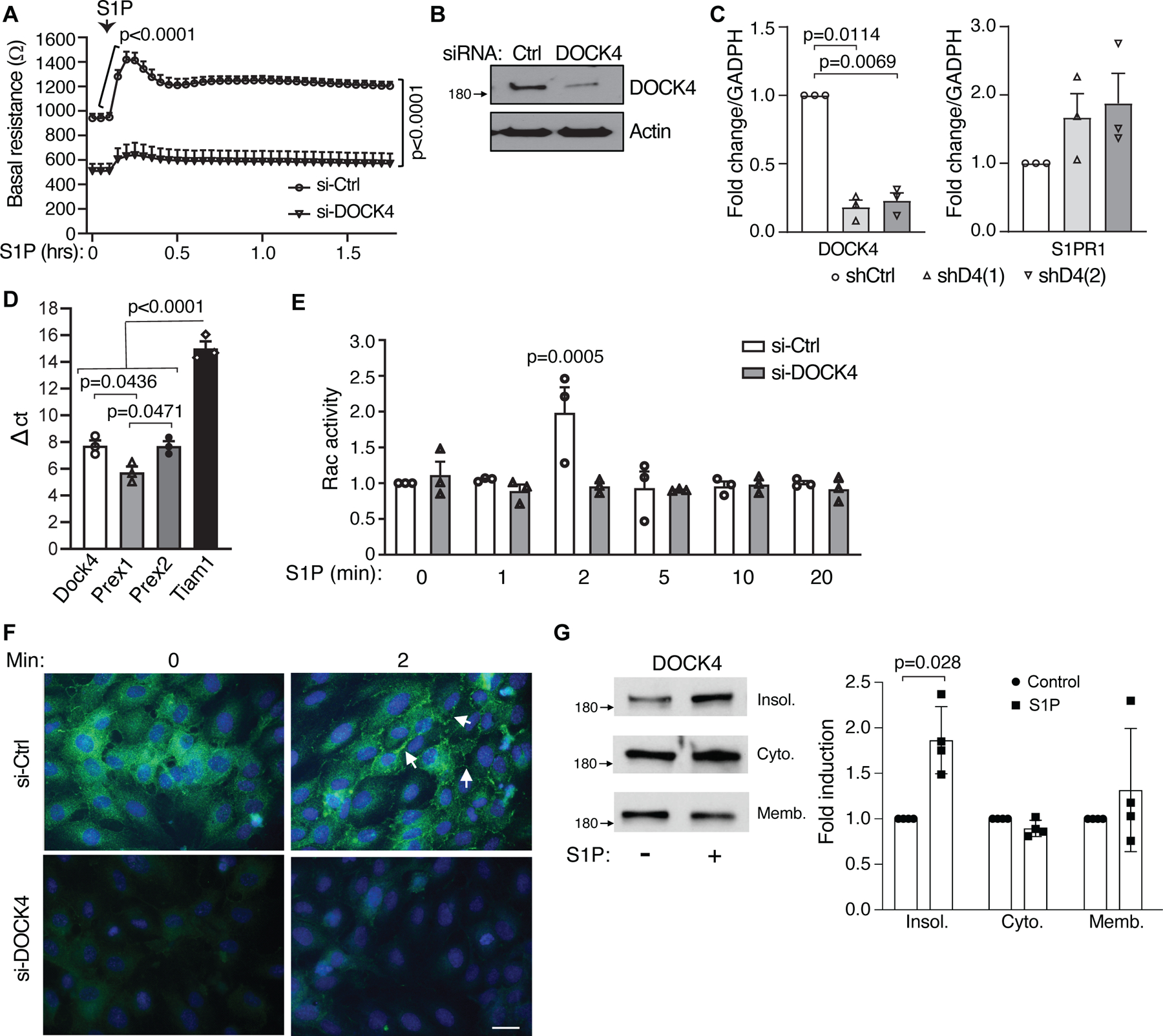
Figure 3. DOCK4 silencing in human lung endothelial cells increases basal permeability.

A. HPAEC were electroporated with control (Ctrl) or DOCK4 siRNA and seeded on gold-plated electrodes for 48 hrs. Cells were serum starved for 2 h after which endothelial barrier function was determined by measuring transendothelial electrical resistance (TEER) over time (in hours) before and after addition of 1 µM S1P. Data are mean ± SEM; Two-way ANOVA multiple comparison (Bonferroni). B. Western blot of DOCK4 in siRNA cells. C. mRNA levels of S1PR1 (left) and DOCK4 (right) in DOCK4 knockdown HPAEC using two independent DOCK4 shRNAs. Data are mean fold increase versus levels in HPAEC transduced with shRNA control lentivirus (Ctrl); Welch’s t test. D. mRNA levels of indicated guanine exchange factors in HPAEC. Data are mean ± SEM.; one-way ANOVA, Tukey’s multiple comparisons. E. HPAEC electroporated with Ctrl or DOCK4 siRNA were plated on 6 well plate. After 48 hrs, cells were starved for 5 hrs, stimulated with 1µM S1P for different times and lysates were used to assess Rac1 activity. Data are mean ± SEM; two-way ANOVA, Tukey’s multiple comparisons. F. HPAEC transfected with Ctrl and DOCK4 siRNA were plated on glass coverslips for 48hrs. Cells were serum starved for 4 hrs followed by treatment with 1 µM S1P for the indicated times in minutes. Cells were fixed and stained for DOCK4 (Green) (n=3). Bar = 20 µm. G. HPAEC were treated with S1P or vehicle control for 1 min and DOCK4 levels in equal amounts of protein from each of the obtained fractions (cytosolic, membrane and insoluble cytoskeletal/nuclear) were analyzed by western blot (left). Western blot quantification of DOCK4 levels after S1P treatment (right). Data are mean fold increase in S1P treated vs. control cells ± SD; Mann-Whitney test.
