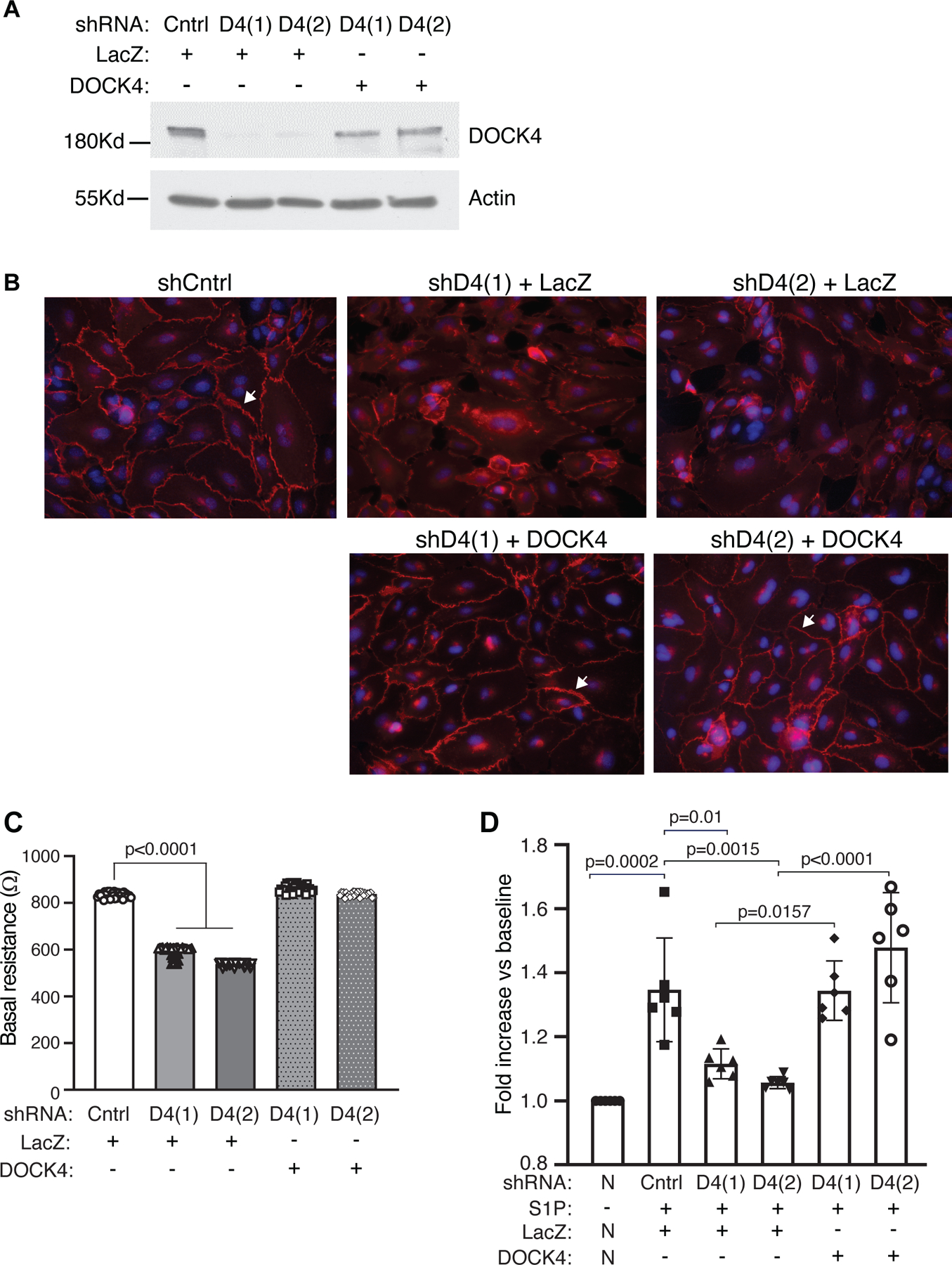
Figure 5. DOCK4 expression in DOCK4 depleted endothelial cells restores barrier function.

A. Transduction of Control (Cntrl) or D4 shRNA endothelial monolayers with wild-type DOCK4 or LacZ adenovirus as indicated. Monolayers transduced with DOCK4 resulted in DOCK4 protein expression. Representative western blot is shown B. VE-cadherin staining (arrow) of Cntrl and D4 shRNA cells transduced with LacZ or DOCK4 adenovirus as indicated. C. HPAEC were transduced with Cntrl or DOCK4 shRNA clones for 48hrs. Cells were trypsinized and plated on electrodes to allow monolayers to form. Wells were then transduced with LacZ or DOCK4 adenovirus for 24hrs. Basal resistance was measured after 72hrs. Data are mean ± SEM; Two-way ANOVA with Tukey’s multiple comparisons. D. Cntrl or DOCK4 shRNA cells, as in Fig. 5C, were transduced with LacZ or DOCK4 adenovirus respectively. After 24hrs, cells were starved for 2 hrs and TEER was examined before and after stimulation with 1 µM S1P. Each experimental sample was normalized (N) to its own baseline (-S1P). Data are mean ± SEM; Two-way ANOVA with Tukey’s multiple comparisons.
