Figure 1.
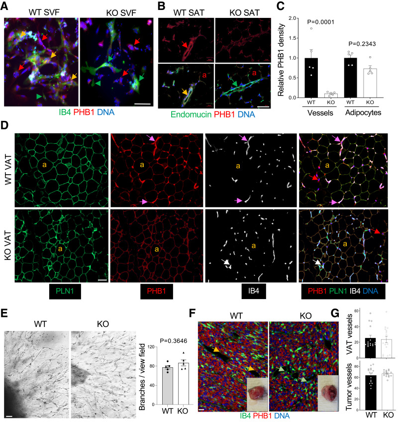
PHB1 EC-KO mice have normal AT and vascularization. A: IF of AT-derived SVF plated in culture. ECs, bound by isolectin B4 (IB4) are PHB1+ in WT mice (yellow arrows) and PHB1− in KO mice (green arrows). Arrowhead: IB4+ monocyte. Red arrows: stromal cells PHB1+ in both WT and KO mice. B: IF on SAT sections reveals a lack of PHB1 in endomucin+ endothelium of KO mice (yellow arrow: colocalization in WT mice); a: PHB1+ adipocytes. Quantification of PHB1 signal in IB4+ endothelium (C) and in perilipin-1 (PLN1)+ adipocytes (D) by ImageJ in IF images. P value by Student t test. D: IF on VAT sections reveals the lack of PHB1 in IB4+ endothelium (purple vs. white arrows); a: PLN1+ adipocytes, which still express PHB1 in KO mice. E: AT explant assay showing that angiogenic sprouting is comparable for WT and PHB1 KO-ECs. Graph: branches/view field in (E) quantified by ImageJ. P value: Student t test. F: Sections of RM1 tumors grown subcutaneously subjected to anti-PHB1 IF show comparable vascular networks stained with IB4 and the lack of PHB1 in ECs of KO mice. Inset images: representative tumors upon resection. G: Quantification of IB4+ vessel density in VAT of mice fed the HFD for 3 months and in tumors (IF images in F by ImageJ). In all mouse experiments, 15-month-old males (n > 3) were used. In all panels, scale bar: 50 µm.

