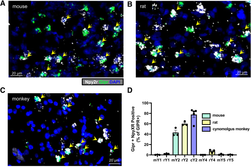
Figure 5.
Specific colocalization of Gipr with Npy2R in mouse, rat, and monkey. Dual-label fluorescence in situ hybridization showing colocalization of Gipr (green) and Npy2r (white) mRNA transcripts in sections containing the AP from mouse (A), rat (B), and cynomolgus monkey (C). Nuclear staining (DAPI, blue) was used to identify brain regions. Yellow arrows indicate Gipr and Npy2r double-labeled cells. Colocalization of Gipr with Npy1, 2, 4, and 5 was assessed in sections from mouse and rat (n = 3) and Gipr with Npy2r in sections from cynomolgus monkey (5 sections from n = 2 monkeys). The percent colocalization (mean and SEM) of Gipr with mouse, rat or cynomolgus monkey NPY1R, NPY2R, NPY4R, and NPY5R (mY1, rY1, mY2, rY2, cY2, mY4, rY4, mY5, rY5).

