Figure 8.
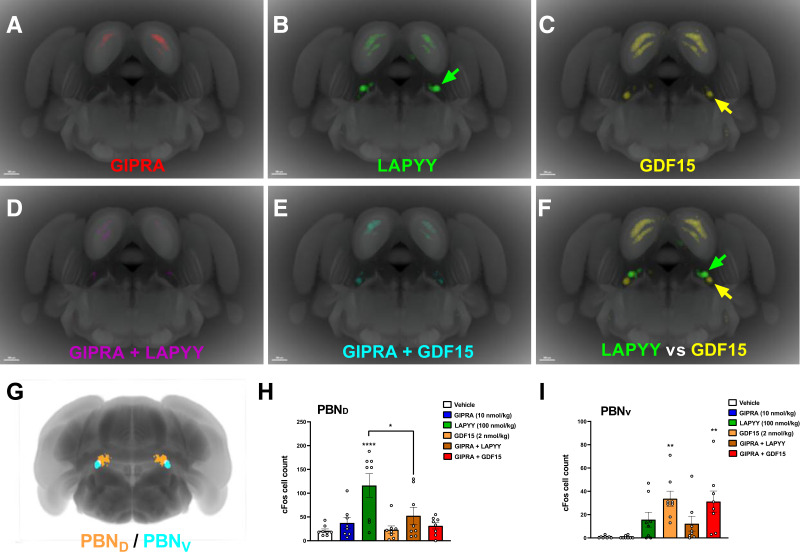
PBND is specifically activated by LAPYY analog and inhibited by GIPRA. Averaged group view (n = 8) of cFos activity at coronal section bregma −5 mm from mice dosed subcutaneously with GIPRA alone (A), LAPYY analog alone (B), GDF15 alone (C), GIPRA + LAPYY analog (D), and GIPRA + GDF15 (E). (F) Virtual overlay of average cFos signal induced by LAPYY in green and by GDF15 in yellow. Note that LAPYY analog and GDF15 activated dorsal-medial (PBND) and ventral-lateral (PBNV) part of the PBN, respectively. G: A 100-μm-thick three-dimensional virtual brain slice with PBND shown in orange and PBNV in cyan. (See research design and methods for details.) H: cFos cell count in three-dimensional PBND. I: cFos cell count in three-dimensional PBNV. Scale bars in panel A–F: 500 μm. Values in H and I are means ± SEM. Statistical analyses included one-way ANOVA followed by Dunnet multiple comparisons test. *P < 0.05; **P < 0.005; ****P < 0.0001.

