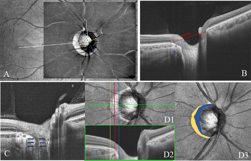
Figure 1.
ONH and peripapillary structural parameter measurement. (A) Measurement of the optic disc tilt ratio and rotation degree by infrared fundus photography on OCT. The optic disc tilt was evaluated as the ratio between the longest and shortest diameters of the optic disc. The rotation was defined as the deviation of the long axis of the optic disc from the reference line (θ), which is 90° from a horizontal line connecting the fovea and the center of the optic disc. (B) ONH parameter measurement in the B-scan image. The BMO distance was evaluated between both sides of BMO. The BL was measured as the straight-line distance between the BMO point and the BT and scleral end. The BT angle (BTA) was defined as the angle between the BMO reference plane and the BT. (C) B-scan OCT image demonstrating focal LC defect (blue arrows). (D) Determination of the presence and area of β-zone and γ-zone PPA. The yellow, red, and blue lines present the boundaries of the RPE, Bruch's membrane, and optic disc margin, respectively (D1, D2). The β-zone PPA is marked in yellow and the γ-zone PPA is marked in blue (D3).

