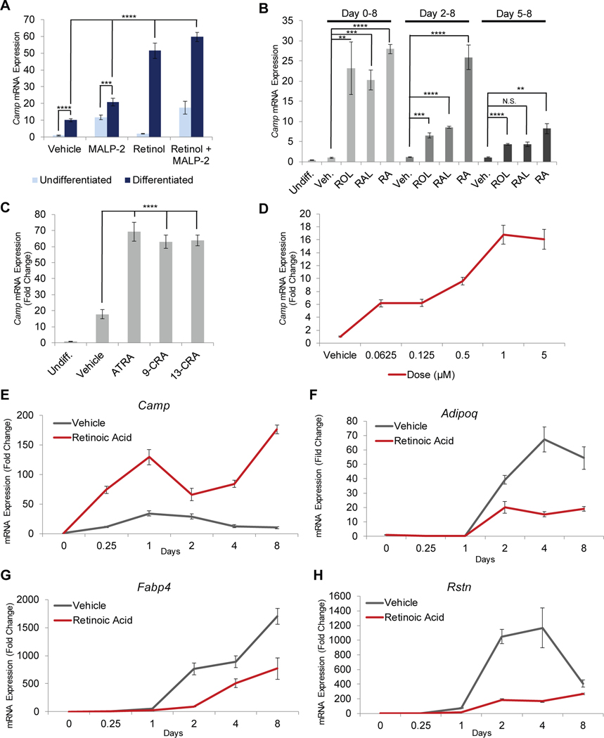
Figure 1. Retinoids significantly increase cathelicidin expression during adipogenesis.
3T3-L1 preadipocytes were differentiated in the presence of retinoids or control, and relative mRNA expression of Camp (A-E), Adipoq (F), Fabp4 (G), or Rstn (H) was assayed by RT-qPCR. (A) 3T3-L1 cells were treated for 24 h in the presence of 1 μM retinol and/or 100 ng/ml MALP-2 or vehicle control in the absence or presence of differentiation media (DM). (B) Cells were differentiated for 8 days and co-treatment was initiated on day 0, day 2, or day 5 in the presence of 1 μM retinol (ROL), all-trans retinal (RAL), retinoic acid (RA), or control. (C) Cells were differentiated for 24 h in the presence of ATRA, 9-cis retinoic acid (9-CRA), or 13-cis retinoic acid (13-CRA). (D) Cells were differentiated for 24 h in the presence of increasing concentrations of RA. (E-H) Cells were differentiated for 8 days in the presence of RA or vehicle control and mRNA expression was assayed at given timepoints. All data are mean ± S.D, *P<0.05, **P<0.01, ***P<0.001, ****P<0.0001 using a Student’s paired T test.

