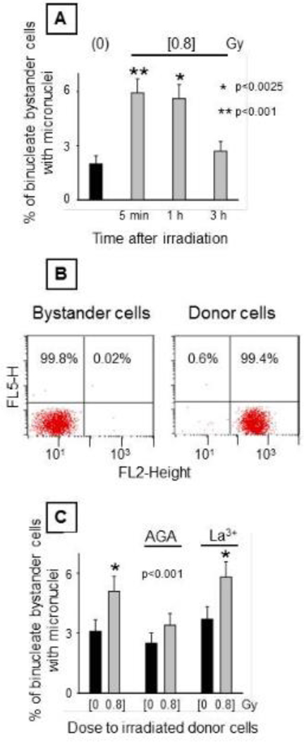
Figure 3. Biological changes in AG1522 bystander cells when co-cultured with irradiated cells.
Donor control or irradiated AG1522 cells exposed to mean absorbed dose of 80 cGy from 3.7 MeV α particles were seeded on the top side of permeable microporous membrane inserts with bystander AG1522 cells growing on their underside. After 5 h of co-culture, the bystander cells were collected. [A] Effect of elapsed time between irradiation of donor cells and co-culture with bystander cells on formation of micronuclei in bystander cells. [B] Flow cytometric analyses of non-dyed bystander cells co-cultured with dyed donor cells. AG1522 donor cells labeled with CellTracker Orange and Calcein were seeded on the top side of the inserts containing non-dyed bystander cells on the bottom side. After 5 h of co-culture, the cells were collected and submitted to Flow Cytometry to determine purity. [C] Effect of inhibitors of connexin channels and hemi channels (50 μM AGA) and hemi channels (100 μM La3+) during 5 h co-culture of bystander cells with irradiated donor cells on the induction of micronuclei in bystander cells.

