
FIGURE 4.
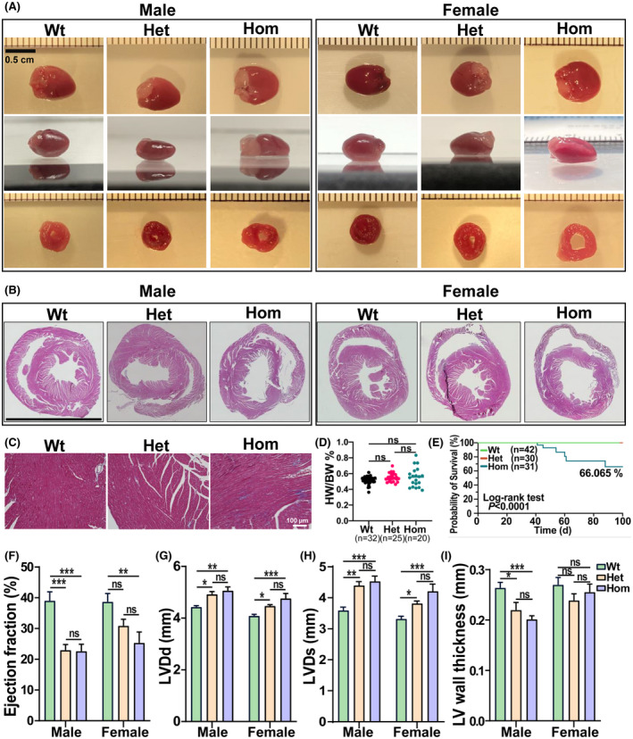
Histology and in vivo cardiac functional assessment of Rbm20S637A KI mice. (A) Gross anatomy of the heart with upper and side views, and slice anatomy at the midventricular level of Wt, Het, and Hom hearts in male and female mice. Bar = 0.5 cm. (B) Representative images of hematoxylin and eosin staining of cross section of mouse hearts at the age of 8 weeks in both male and female mice. Bar = 0.5 cm. (C) Representative images of Masson's trichrome staining in male mice at 8 weeks old. Bar = 100 µm. (D) Heart weight versus body weight at the age of 8 weeks in both male and female mice (n = 32 in Wt, 25 in Het, and 20 in Hom). (E) Kaplan–Meier survival curves of Wt, Het and Hom male and female mice. Statistical significance was determined by log‐rank (Mantel–Cox) test with two degrees of freedom (n = 42 in Wt, 30 in Het, and 31 in Hom). (F) Ejection fraction (EF), (G) left ventricular diastolic diameter (LVDd), (H) left ventricular systolic diameter (LVDs), and (I) left ventricular wall thickness (measured by 2*LVPW(d)/LVDd; mm) from echocardiographic imaging of mice at the age of 2 months (n = 7 males and 10 females in Wt, n = 6 males and 7 females in Het, and n = 8 males and 7 females). Mean ± SEM. Ns, not significance, *p < .05, **p < .001, and ***p < .0001