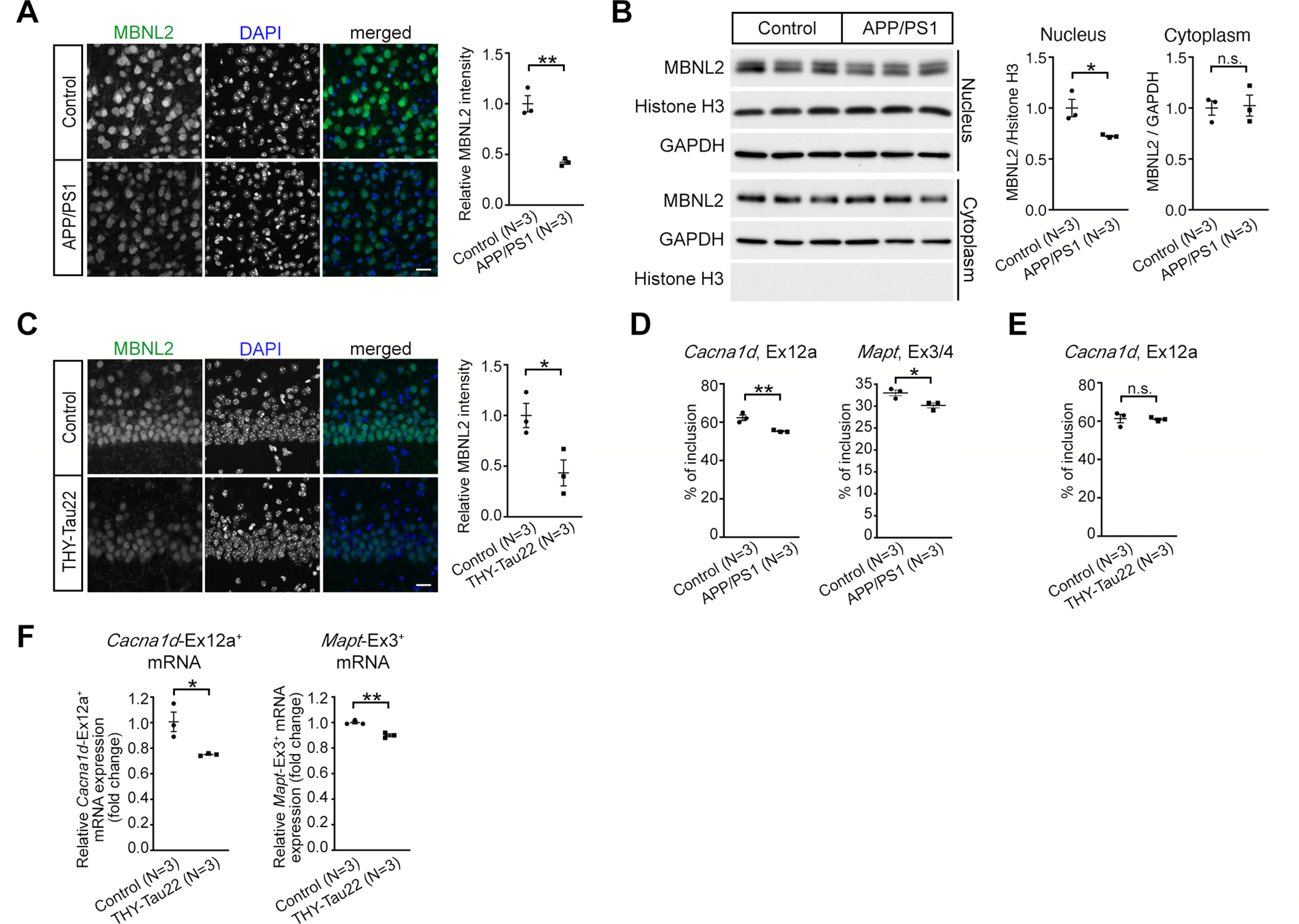
Figure 2.

Neurodegeneration-reduced MBNL2 expression and aberrant MBNL2-reuglated splicing are detected in the mouse brains for AD. A, The intensity of MBNL2 immunoreactivity was reduced in cortical Layer V neurons of female APP/PS1 mice at age 12 months (p = 0.0022). Non-Tg animals at the same age were used as controls. B, Western blot analysis of MBNL2 expression in the nuclear and cytoplasmic fraction from the cortical region of the control (non-Tg) and APP/PS1 mice at age 10 months. Relative MBNL2 level in the nuclear and cytoplasmic fraction, normalized with Histone H3 and GAPDH, respectively, was quantified (Nucleus: p = 0.0322; Cytoplasm: p = 0.8609). C, Immunofluorescent staining of MBNL2 expression in the caudal CA1 neurons of female non-Tg (Control) and THY-Tau22 mice at age 15 months (p = 0.0318). D, The percentage of inclusion of Cacna1d exon 12a (p = 0.0093) and Mapt exons 3/4 (p = 0.0295) in the cortical tissues of APP/PS1 mice. E, The percentage of inclusion of Cacna1d exon 12a using RNA collected from the entire hippocampal tissues of THY-Tau22 mice (p = 0.8899). F, Quantification of the levels of exon 12a-containing Cacna1d (Cacna1d-Ex12a+) and exon 3-containing Mapt (Mapt-Ex3+) mRNA by RT-qPCR after normalization to Gapdh level using RNA collected from the CA1-enriched tissues of THY-Tau22 mice (Cacna1d-Ex12a+: p = 0.0284; Mapt-Ex3+: p = 0.0028). Number of animals (N) in each group is indicated. Data are mean ± SEM; *p < 0.05, **p < 0.01, by Student t test. n.s., not significant. Scale bar: 20 µm (A and C).
