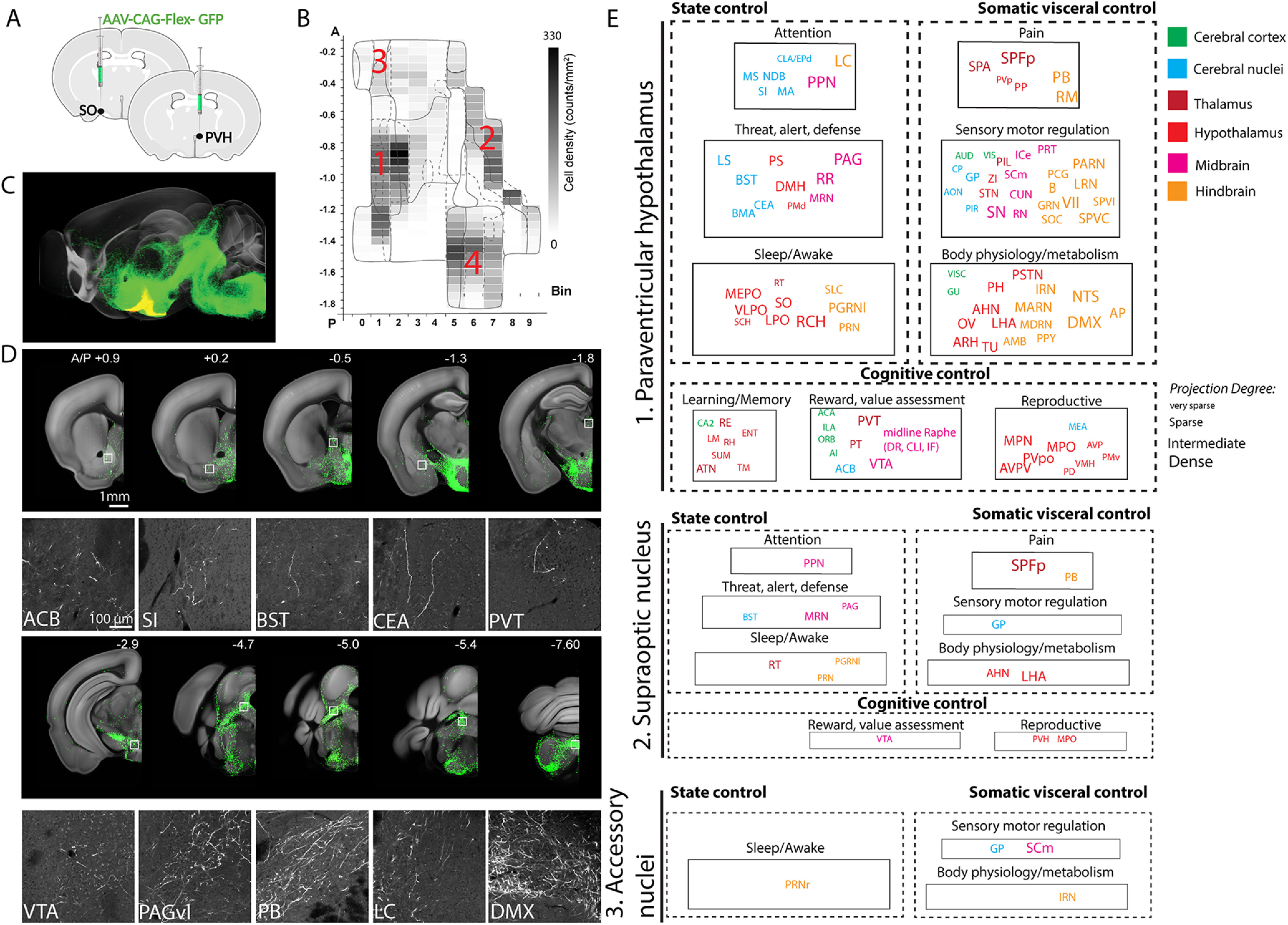
Figure 2.

Anterograde projection map of Oxt neurons. A, Conditional AAV-GFP was injected in Oxt neuron containing hypothalamic areas. B, Four major areas of viral injections, 1: the PVH, 2: the SO, 3: the AN, 4: the TU area. C, Projection outputs from the PVH (green) and SO (yellow) Oxt neurons registered in the Allen CCF. See also Movie 2. D, Examples of long-range projections (green) from Oxt neurons in the PVH. The bottom panel is high mag images from white boxed areas in the top panel. E, Nine functional circuits that receive long-range projection from Oxt neurons in the three different injection area 1–3. Color and size of each ROI represent anatomic ontology and the abundance (degree) of the projection. The full name of abbreviations can be found in Table 2.
