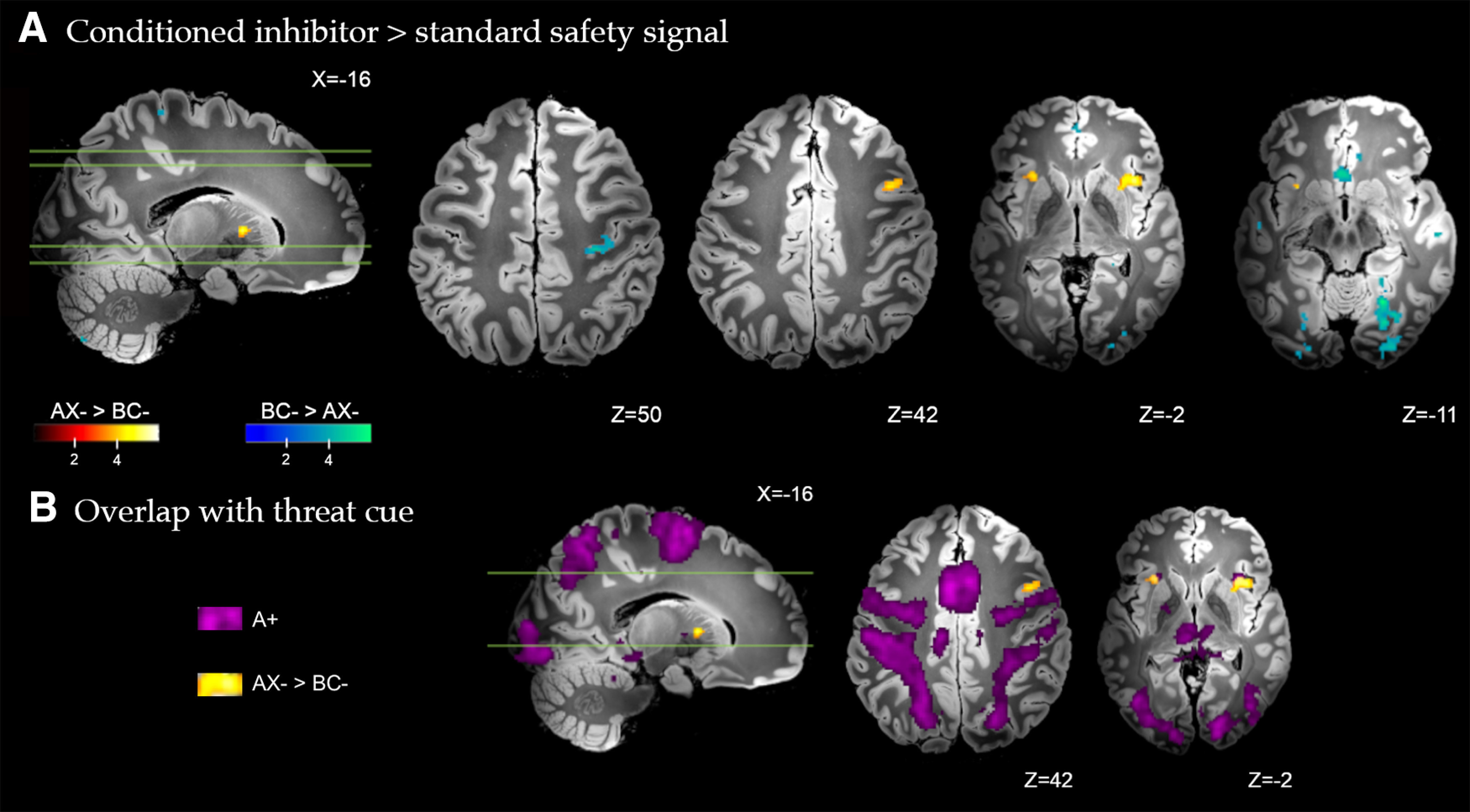
Figure 3.

A, Brain regions with significant differential response to the conditioned inhibitor (AX– > BC–) versus the standard safety signal (BC– > AX–) across all trials of conditioning. Whole-brain FDR-corrected (p < 0.05) results are displayed on a high-resolution anatomic template in MNI space. B, Partial overlap between the differential response to the conditioned inhibitor versus standard safety signal (AX– > BC–, yellow), and the response to the conditioned threat alone (A+, purple) in activation of the anterior insular cortex.
