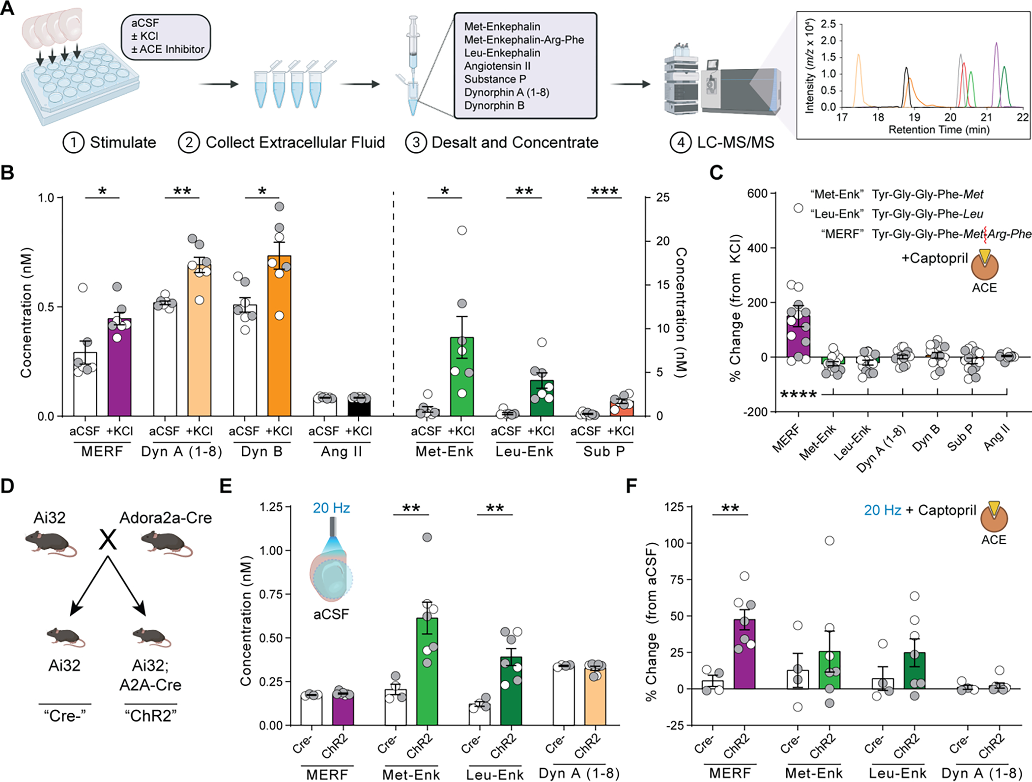
Fig. 2. ACE selectively degrades MERF in the extracellular space.

(A) Quantification of neuropeptide release from brain slices using LC-MS/MS. (B) Extracellular neuropeptide levels from slices submerged in normal aCSF or 50 mM KCl. (C) Percent change in extracellular neuropeptide levels after KCl stimulation in presence versus absence of captopril (10 μM). Inset shows enkephalin amino acid sequences and site of enzymatic cleavage of MERF by ACE (red line). (D) Breeding strategy to generate mice expressing channelrhodopsin-2 in D2-MSNs. (E) Extracellular neuropeptide levels from slices following optogenetic stimulation at 20 Hz. (F) Percent change in extracellular neuropeptide levels after optogenetic stimulation in presence versus absence of captopril (10 μM). Data are mean ± s.e.m. for all panels; open and closed circles indicate samples from female and male mice, respectively. *P<0.05, **P<0.01, ***P<0.001, ****P<0.0001, ANOVA followed by simple effect test (B, E, F) or Fisher’s LSD post-hoc test (C); see Data S1 for complete statistics.
