
Figure 2.
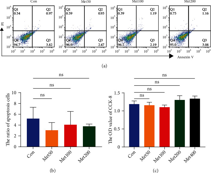
Metformin did not influence the proliferation of UC-MSCs in vitro. (a, b) Representative graphs and statistical analysis of Annexin V/PI double staining were displayed. (c) The OD value of CCK-8 after different concentrations of metformin (0, 50, 100, 200, and 400 μM) for 48 h. Data were expressed as mean ± SD from three independent experiments. Met: metformin; ns: no significance.