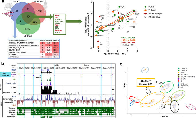
Fig. 6. An evolutionary conserved StemLeish gene signature recapitulates cross-species epigenetic and transcriptional signatures of visceral leishmaniasis.
a In vivo validation of StemLeish signature in three independent cohorts of human VL patients from Brazil, from India and HIV-VL co-infected patients from Ethiopia, all fold-changes of DEG from untreated patients with active disease vs. post-treatment blood samples. Venn diagram (upper left panel) shows overlap between murine and human signatures. Human Phenotype Ontology (MSigDb) enrichment analysis indicates the StemLeish signature phenocopies the complete clinical picture of human VL (lower left panel). Significant correlations (Spearman) between StemLeish and human VL DEG in all three cohorts (right panel). b Integrated epigenetic, transcriptomic and genomic analysis of the Rgs1 locus, visualized in UCSC Gene browser. Open chromatin regions (DNase I Hypersensitive Sites) in purified CD34+ HSC (indicated as CD34+) and HSC undergoing myeloid differentiation (indicated as CD34−) are indicated by light blue shades. Transcriptomic data (RNAseq) represent mapped reads present in purified CD14+ vs. purified mobilized CD34+ cells, with embryonic stem cells (ESC) and lymph node RNAseq data plotted for comparison. Genomic data show conservation score across 100 different vertebrate species (blue: conserved, brown: not conserved), with leishmaniasis and tuberculosis model species (rhesus macaques, mouse, hamster, dog, zebrafish) plotted individually (green panels below), as compared to the human reference genome. c Reanalysis of single-cell RNAseq data50 using UMAP (Unifold Mapping) revealed presence within purified human CD34+ cells of an RGS1high phenotype in several progenitor subsets [Erythro-Myeloid Progenitors (EMP), Lymphoid/Myeloid Progenitors (LMPP7), HSC in a metabolically active state (HSC/Met. state)]. Arrows demonstrate clusters containing RGS1high cells (orange vs. gray RGS1low cells). Arrow sizes are proportional to the frequency of RGS1high cells. Clipart used in diagram (a) was obtained from Servier Medical Art (https://smart.servier.com).

