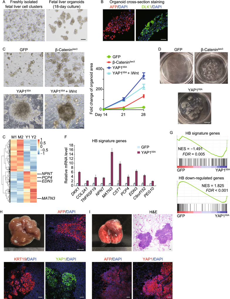
Figure 1.
Modeling hepatoblastoma development with human fetal liver organoids reveals YAP1 activation is sufficient for tumorigenesis. (A) Brightfield images of freshly isolated human fetal liver cell clusters and 18-day cultured organoids. Scale bar, 200 μm. (B) Immunofluorescence staining for the hepatoblast markers AFP and DLK1. Scale bar, 50 μm. (C) Brightfield images of 21-day cultured fetal liver organoids transfected with lentiviral GFP, β-cateninΔex3 or YAP15SA. Growth rate of organoids were quantified. iWnt: Wnt inhibition by 5 μmol/L IWP-2 treatment. Scale bar, 200 μm. (D) Organoids transfected with indicated genes were cultured for 60 days. Scale bar, 250 mm. (E) mRNA expression heatmap of differentially expressed genes for Mock (M1, M2) and YAP1-activated (Y1, Y2) organoids. (F) GFP and YAP1-transfected organoids were harvested to examine the expression of HB signature genes using qRT-PCR. H3 was used as an internal control. Data were presented as means ± SD (n = 3). (G) GSEA enrichment analysis of Mock versus YAP1-activated organoids for HB/normal top 200 genes (top) and HB/normal last 200 genes (bottom). (H and I) YAP1-activated HB organoids were transplanted into the liver capsule of NSG mice. Tumors in liver (H) and metastatic foci in lung (I) were subjected to immunofluorescence staining for AFP, KRT19 and YAP1. Scale bar, 100 μm

