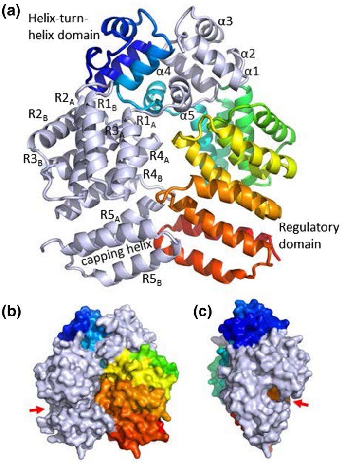
FIGURE 1.

Crystal structure of Rgg144. (a) The Rgg homodimer. Polypeptides are light gray or rainbow colored, with the N‐terminus in blue and the C‐terminus in red. Helices have been numbered according to Parashar et al. (2015). The DNA‐binding helix is α3. (b) and (c) surface representation of the front and side views of the Rgg homodimer. The position of the putative peptide‐binding groove is shown by the red arrow.
