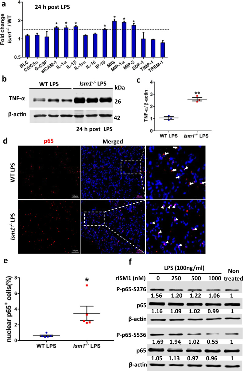
Fig. 7.
ISM1 deficiency increased proinflammatory cytokine/chemokine production in lung and promoted LPS-induced NF-κB activation. a Lung cytokine/chemokine profile determined by an inflammatory cytokine antibody array using lung homogenates at 24 h post LPS challenge. * represents p < 0.05. n = 4 mice per group, 1 total lung lysate per mouse. b, c Increased levels of TNF-α in Ism1−/− lung 24 h post LPS challenge shown by Western blot of whole lung lysate. ** represents p < 0.01. n = 3 mice per group, 1 total lung lysate per mouse. d, e IF staining showing increased p65 NF-κB nuclear translocation in Ism1−/− lung. Representative images showing increased signal co-localization of the p65 NF-κB (red) and the DAPI nucleus (blue) in the lung sections of Ism1−/− mice d and the quantification of nuclear p65 NF-κB in panel d, showing the percentage of cells with nuclear localized p65 in WT and Ism1−/− lung (e). n = 5 mice per group, 2 sections per lung, 5 microscopic fields per section. f rISM1 dose-dependently suppresses LPS-induced NF-κB p65 phosphorylation at S276 and S536 in cultured mouse MH-S alveolar macrophage cells. Cells are harvested at 30 min post LPS treatment (100 ng/ml). Based on two independent experiments

