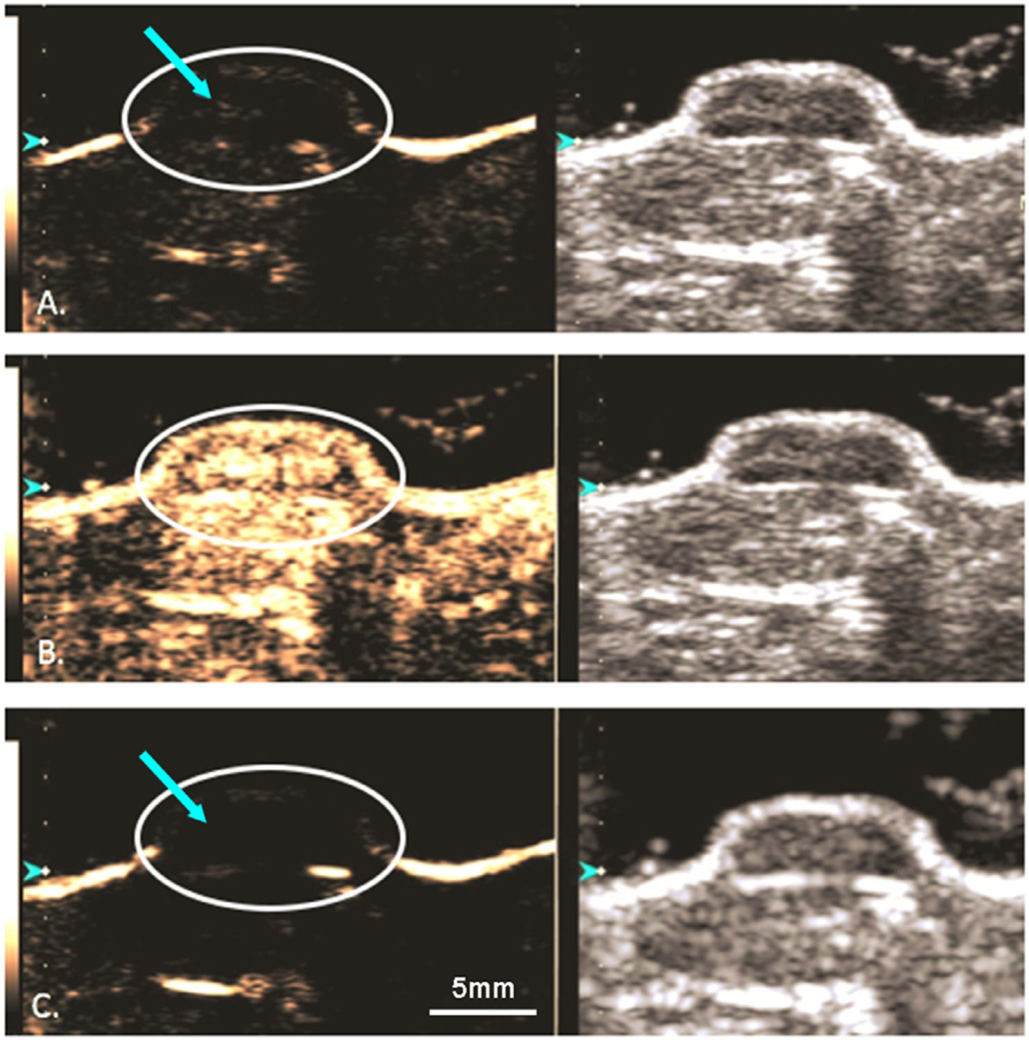
Fig. 7.

Dual ultrasound of a contrast enhanced (left) and B-mode (right) human PDAC xenograft in a mouse model, MIA PaCa-2. A) GEM-MB are visualized within the tumor (arrow). B) A 4 second, 1.35 mechanical index destructive pulse was delivered to the tumor. C) Post destructive pulse displaying an absence of microbubbles within the tumor. Scale bar: 5mm.
