
Fig. 3. Endymocyte cell type family.
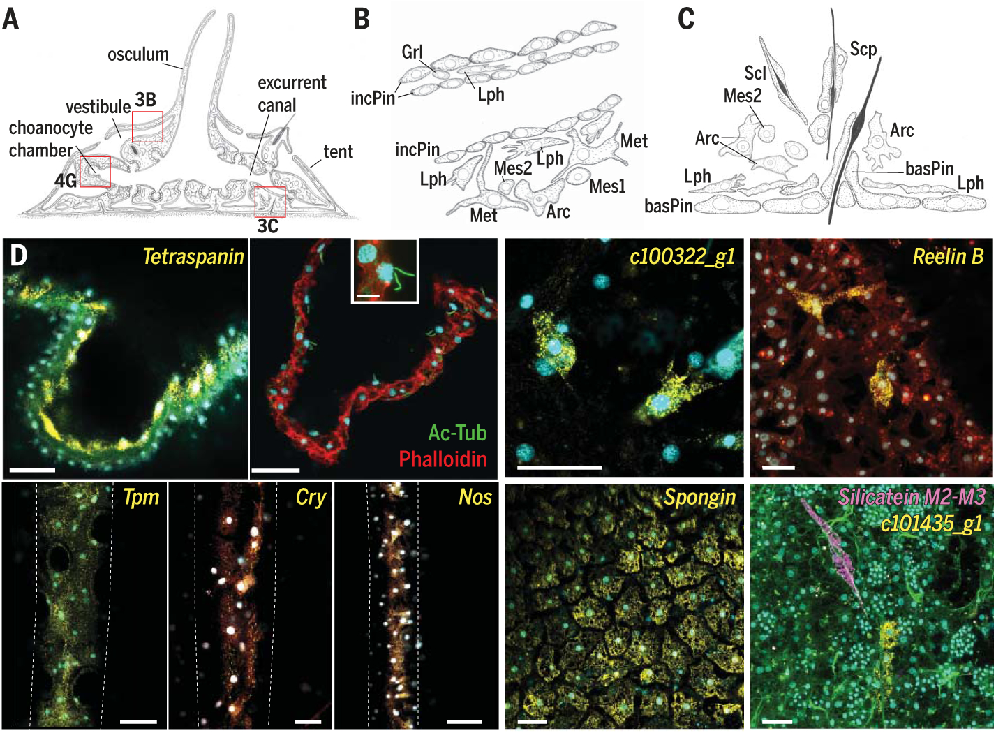
(A) Illustration of juvenile S. lacustris. Red boxes outline locations illustrated in other figure panels. (B) Drawing of incurrent pinacocytes (incPin) that make up the tent and vestibule, with adjacent mesenchymal cells: lophocytes (Lph), metabolocytes (Met), archaeocytes (Arc), mesocytes 1 (Mes1), and mesocytes 2 (Mes2). (C) Illustration of spicule production (sclerocytes, Scl), transport (sclerophorocytes, Scp), and anchoring (basopinacocytes, basPin). (D) smFISH of endymocyte markers. incPin, Tpm; apnPin, Tetraspanin; lph, c100322_g1; met, Reelin B; basPin, Spongin; Scp, c101435_g1. Membrane stains Fm-143Fx (red) and CellBrite Fix (green; also possibly stains collagen); nuclei 4′,6-diamidino-2-phenylindole (DAPI) stain (cyan). Scale bar, 30 μm.