
Fig. 4. Peptidocyte and amoeboid-neuroid cell type families.
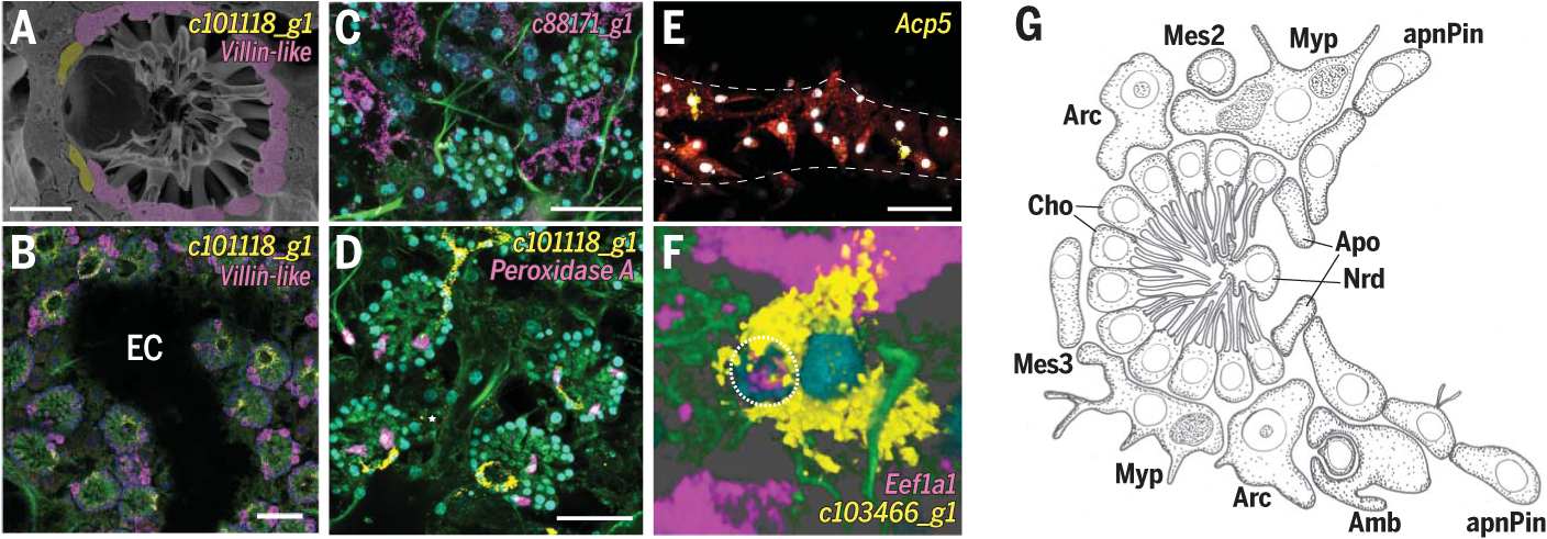
(A) SEM of choanocyte chamber. Scale bar, 10 mm. (B to F) smFISH of neuroid and amoeboid markers: choanocytes (Cho), villin-like; apopylar cells (Apo), c101118_g1; myopeptidocytes (Myp), c88171_g1; neuroid cells (Nrd), Peroxidase A; granulocytes (Grl), Acp5; and amoebocytes (Amb), c103466_g1. Dashed line indicates epithelial tent. Dotted line outlines archaeocyte being engulfed by amoebocyte. Membrane stains Fm-143Fx (red) and CellBrite Fix (green); nuclei DAPI stain (cyan). Scale bars, 30 μm. EC, excurrent canal. (G) Illustration of choanocyte chamber and neighboring mesenchymal cells.