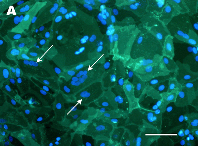
Fig. 3.

Confirmation of syncytialisation by staining of cell membranes. A Fluorescent image of PKH67 staining of cultures of cytotrophoblasts from a placentae of 39 weeks of gestation (isolated using the protocols described in [110]), in which spontaneous syncytiotrophoblast differentiation has occurred over the 30 day culture period. Nuclei are counterstained with Hoechst 33342. Multinuclear clusters within a single membrane (demonstrating syncytiotrophoblast) are indicated with white arrows. Scale bar = 100 μm
