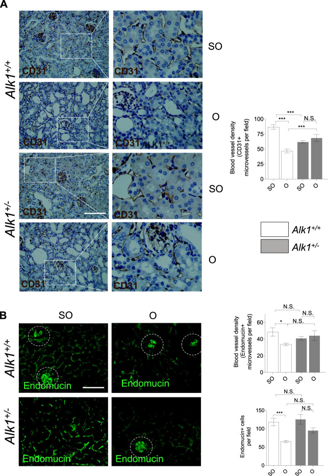
FIGURE 6.
Impaired peritubular capillaries rarefaction in Alk1 +/− mice. (A) CD31 immunostaining in SO and O kidneys from Alk1 +/+ and Alk1 +/− mice and blood vessel density analysis, represented as CD31 + vessels per field. (B) Endomucin immunofluorescence staining in SO and O kidneys from Alk1 +/+ and Alk1 +/− mice and blood vessel density quantification from endomucin staining, represented as microvessels per field (upper graph) and endomucin+ cells per field (lower graph) in SO and O kidneys from Alk1 +/+ and Alk1 +/− mice. *p < 0.05; ***p < 0.001; N.S. Not statistically significant (Two-way ANOVA). Squares in (A) indicate the zoomed areas. Scale bar = 200 microns in both panels. Blood vessels from the glomeruli in (B) (highlighted as cropped áreas) were not counted.

