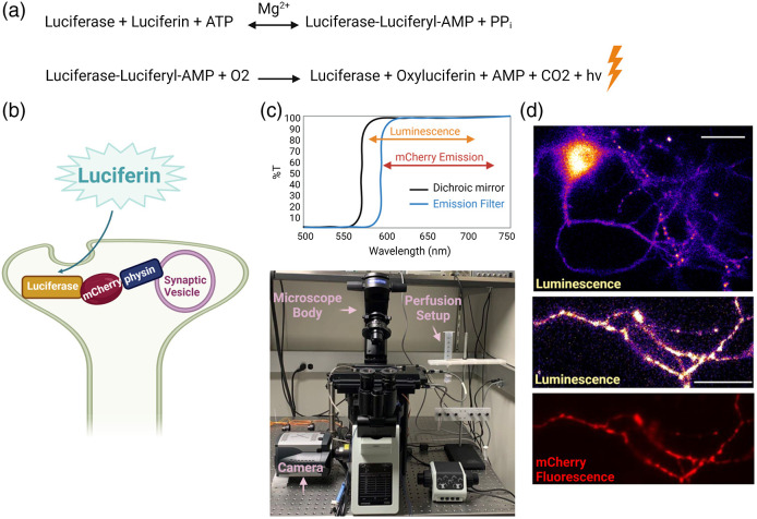
Fig. 1.
Bioluminescence imaging of cytosolic ATP in nerve terminals. (a) The bioluminescence chemical reaction in which the enzyme luciferase uses luciferin and ATP to produce light denoted as . (b) Schematic of a hippocampal nerve terminal expressing Syn-ATP in which luciferase is anchored to synaptic vesicles with synaptophysin (physin) and mCherry is used as an inert fluorophore. (c) An optimized dual fluorescence and luminescence microscopy setup (bottom) where a long-pass 590-nm filter replaces an emission filter to maximize luminescence photon collection (top). (d) Representative luminescence and mCherry fluorescence images of a hippocampal neuron (top) and an axon bearing several nerve terminals (bottom). Scale bar, .

