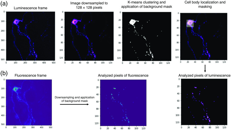
Fig. 2.
An image analysis pipeline for background signal determination and cell body removal. (a) The luminescence image of a neuron was downsampled from to . K-means clustering algorithm was implemented on pixel values to produce two complementary clusters of background and desired signals. A background mask was applied to remove background signals from the image (black and white panel). Next, the region with the highest total signal intensity was detected and deemed as the cell body. Both background and cell body were removed from further analysis. (b) Background and cell body masks generated from the luminescence image were applied to the fluorescence image.

