Fig. 7.
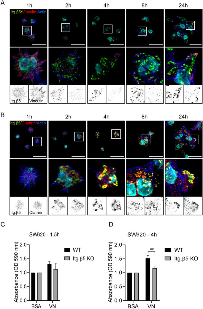
Integrin β5 in clathrin lattices mediates adhesion to vitronectin ∼4 h after cell seeding. (A,B) SW620 cells were seeded on vitronectin-coated coverslips and fixed at the indicated time points. Representative immunofluorescence images showing integrin β5 (green in merge), vinculin (A) or clathrin (B) (red in merge), actin in blue, and the cell nuclei in cyan. Scale bars: 20 μm. Representative images are shown of two independent experiments performed in duplicate. (C,D) Adhesion assay performed 1.5 h (C) and 4 h (D) after seeding SW620 wild-type (WT) and β5 knockout cells on vitronectin (VN) (three biological replicates; each experiment in triplicate; bars show mean±s.d). **P<0.01 (two-sided unpaired Student's t-test).

