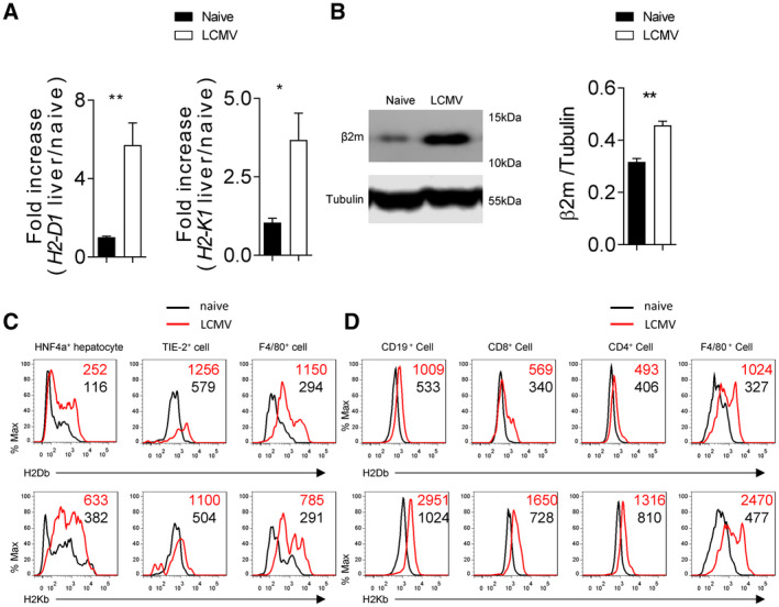
FIG. 2.

H2‐Db is rapidly up‐regulated in liver tissue following LCMV infection. C57BL/6 mice (H2‐b) were infected with 2 × 10⁴ pfu of LCMV Docile. Four days following infection, H2‐D1 and H2‐K1 messenger RNA was quantified by real‐time PCR using RNA isolated from liver tissue (n = 6) (A). (B) Beta‐2 microglobulin protein expression in liver tissue was determined by immunoblot analysis (n = 3). (C) Expression of H2‐Db (upper panels) and H2‐Kb (lower panels) was measured in different cell types of single cell suspended liver tissue from infected and naïve animals by flow cytometry (n = 7 for Kupffer cells and hepatocytes; n = 6 for endothelial cells; average of mean fluorescence intensity (MFI) is listed on the upper‐right corner). (D) Expression of H2‐Db (upper panel) and H2‐Kb (lower panel) was determined by flow cytometry in different immune cells in spleen tissue (n = 7; average of MFI is listed on the upper‐right corner). Error bars show SEM. *P < 0.05 and **P < 0.01 between the indicated groups.
