
FIG. 3.
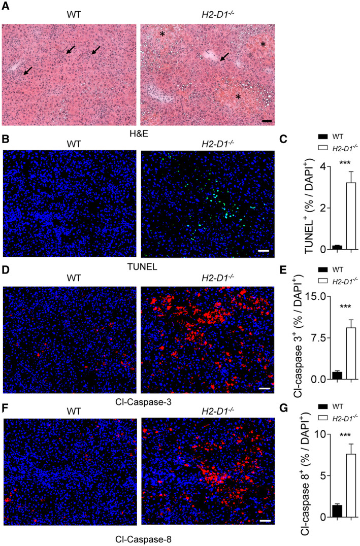
H2‐Db protects liver tissue during LCMV infection. WT and H2‐D1−/− mice were infected with 2 × 10⁴ pfu of LCMV Docile. (A‐I) At day 9 following infection, sections of snap‐frozen liver tissue were analyzed using H&E staining (one representative set of images of n = 5 is shown; scale bar = 50 µm; stars indicate the necrotic areas; arrows indicate the inflammatory infiltrates) (A). Sections of snap‐frozen liver tissue were analyzed for TUNEL (B,C), cleaved Casp3 (D,E), and cleaved Casp8 (F,G); expression was determined and quantified using fluorescent staining of tissue sections. One representative set of images of n = 8 is shown; scale bar = 50 μm; three fields of each section were analyzed for the frequency of TUNEL, cleaved Casp3, or cleaved casp8 positive cells out of the total 4´,6‐diamidino‐2‐phenylindole (DAPI)–positive cells (n = 24). Error bars show SEM; ***P < 0.001 between the indicated groups. Abbreviation: Cl, cleaved.