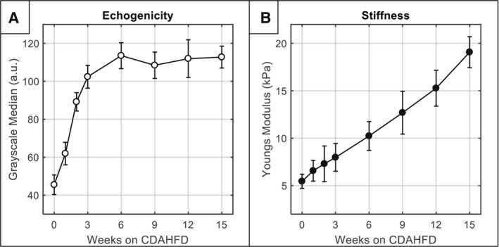
FIG. 4.

Longitudinal progression of noninvasive imaging parameters (mean ± SD). (A) Echogenicity over time measured by GSM. (B) Liver stiffness over time measured by SWE. Note that the full sample size is included at each timepoint, and sample size decreased over time by 5 mice due to sacrifice for histology (e.g., n = 40 mice at week 0, n = 35 mice at week 1, etc.).
