Figure 1.
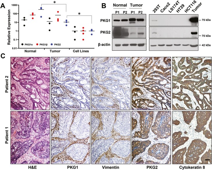
Expression of PKG isoforms in human colon-derived tissues and cancer cell lines. Comparison of PKG isoform expression in patient-derived colon matched normal/tumor specimens and colon cancer cell lines by (A) RT-qPCR and (B) Western blotting. (C) Colectomy specimens were processed for IHC to detect type 1 or 2 PKG as indicated. Serial sections were stained for vimentin and cytokeratin 8 as markers for stroma and tumor epithelium (respectively). Magnification is the same in all panels, scale bar = 100 μm. ∗P < 0.05 using ANOVA with Tukey’s multiple comparison test. PKG expression in the cell lines was less than tumor tissue using two-sided t-tests for each isoform (P < 0.05). IHC, immunohistochemistry.

