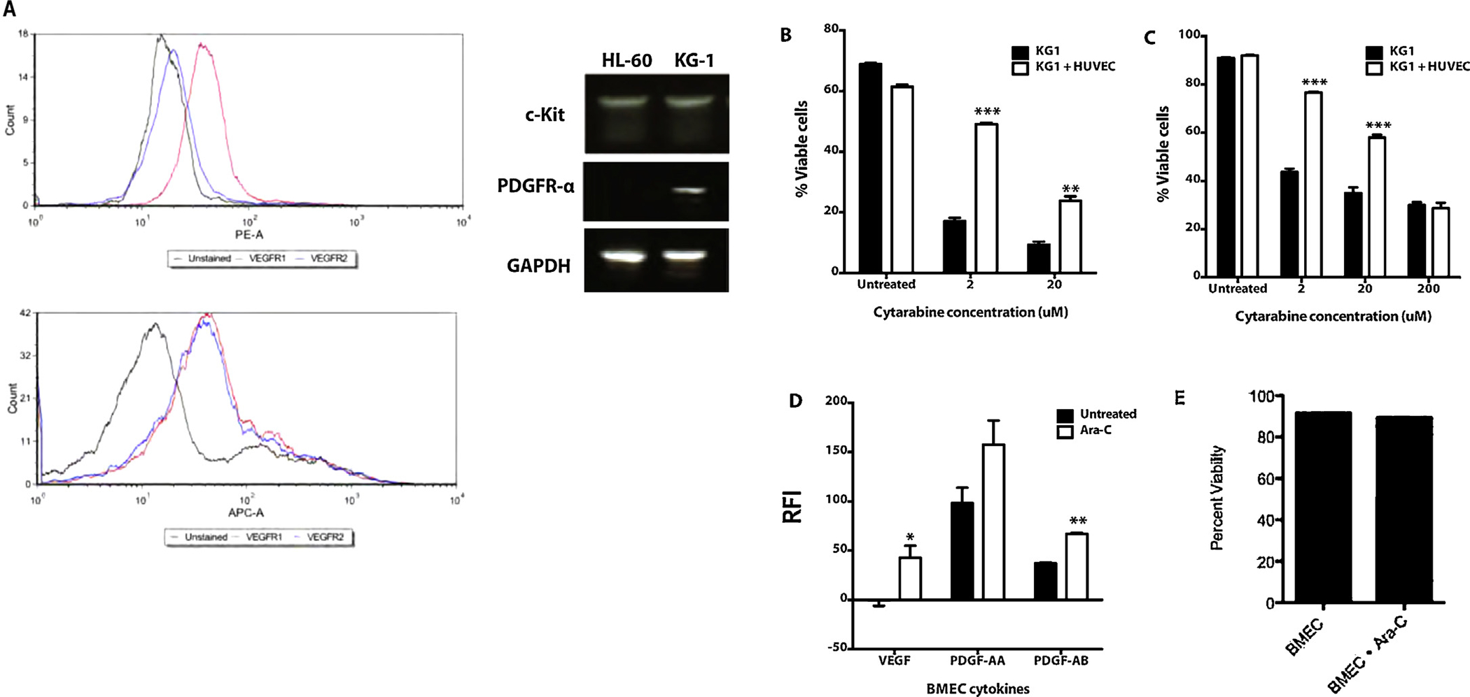
Fig. 1.

Endothelial cells protect leukemia cells from chemotherapy induced apoptosis in co-culture. (A) Flow cytometry shows HL-60 cells express VEGFR1 while KG1 cells express VEGFR1 and VEGFR2. PCR demonstrates HL-60 cells express c-Kit while KG-1 cells express c-Kit and PDGFR-a. (B) KG1 cells were seeded in direct co-culture with HUVECs and treated with varying concentrations of cytarabine. After 48 h of treatment the cells were stained with Annexin V and PI, and then analyzed by flow cytometry. KG1 AML cells were more resistant to cytarabine treatment when in co-culture with ECs (p < 0.001). (C) KG1 cells were seeded in transwell co-culture with HUVECs and treated with varying concentrations of cytarabine. After 48 h of treatment the cells were stained with Annexin V and PI, and then analyzed by flow cytometry. KG1 AML cells were more resistant to cytarabine treatment when in transwell co-culture with ECs (p < 0.001). (D) Angiogenic cytokine secretion from primary human BMECs. After cytarabine chemotherapy, BMECs secreted significantly increased VEGF-A (40-fold, p < 0.05) and PDGF-A/B (1.6-fold, p < 0.005). RFI = Relative Fluorescence Intensity. (E) BMEC viability after 24 h treatment with cytarabine chemotherapy at clinically relevant concentration, 20 μM. Flow cytometry analysis revealed no reduction in viability.
