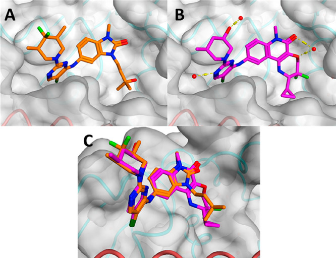
Figure 1.

(A) X-ray structure of the BCL6 BTB domain with the bound ligand CCT369260 (PDB: 6TOM, orange).14 (B) X-ray structure of the BCL6 BTB domain with the bound ligand CCT373566 (PDB: 7QK0, magenta). (C) Overlaid X-ray structures of CCT369260 and CCT373566; the piperidines of both compounds are found to reside in the same position. In all panels, the surface of the BCL6 dimer is shown as a gray transparent surface, with the two individual monomers displayed in ribbons and colored in orange and cyan, respectively. The selected water molecules are shown as red spheres, and H-bonds are shown as yellow dashed lines.
