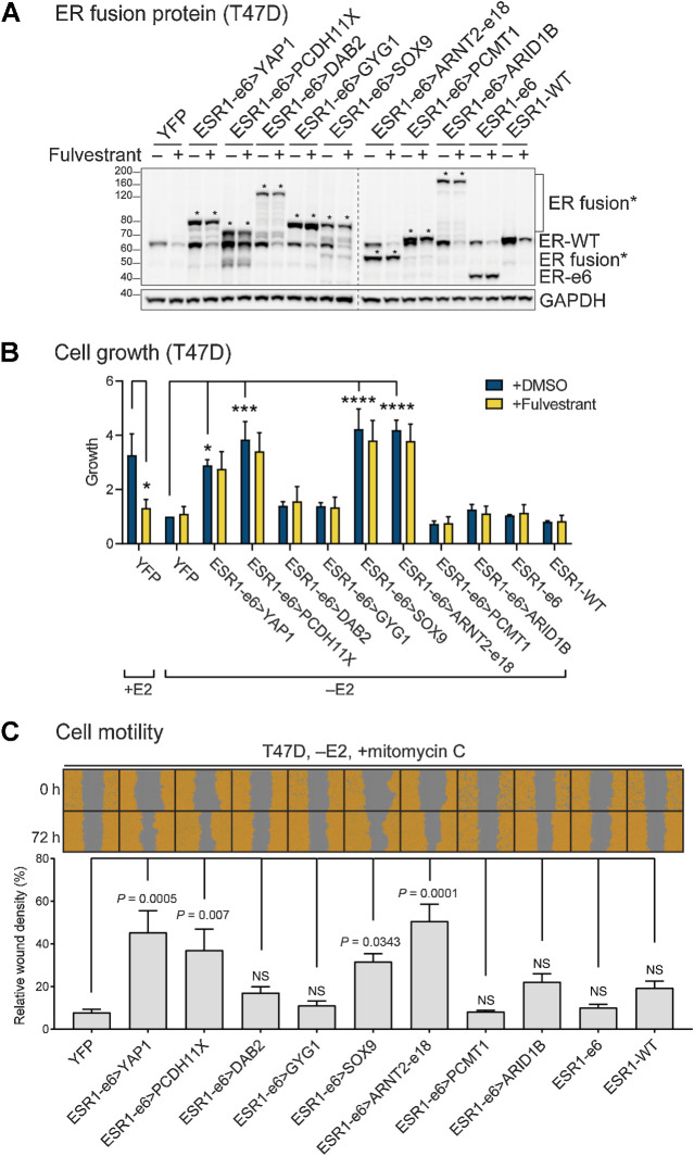
Figure 2.
ESR1 fusion proteins drive ET-resistant growth and promote hormone-independent motility of ER+ breast cancer cells. A, Immunoblotting of ERα and ESR1 fusion proteins with an N-terminal ERα antibody in lysates made from hormone-deprived stable T47D cells. Asterisks indicate ER fusion proteins. GAPDH serves as a loading control. The dashed line indicates two separate blots that were conducted at the same time. The representative image is from three independent experiments. B, Cell growth was assayed in hormone-deprived stable cells (mean ± SEM; n = 3). One-way ANOVA followed by Dunnett multiple comparisons test was used to compare data of hormone-deprived ESR1 fusion expressing cells with YFP control cells in the vehicle (+DMSO) group. Two-way ANOVA followed by Bonferroni test was used for multiple comparisons for each stable cell line after 100 nmol/L fulvestrant treatment in the presence or absence of 10 nmol/L estradiol (E2). *, P < 0.05; ***, P < 0.001; ****, P < 0.0001. See Supplementary Fig. S1B for the complete data. C, Cell motility was detected using scratch wound assays in hormone-deprived stable T47D cells, treated with mitomycin C to block proliferation (mean ± SEM; n = 3). Cells are pseudo-colored orange to aid visualization. One-way ANOVA followed by Dunnett multiple comparisons test was used to compare each stable T47D cell line with YFP control cells. NS, not significant.

