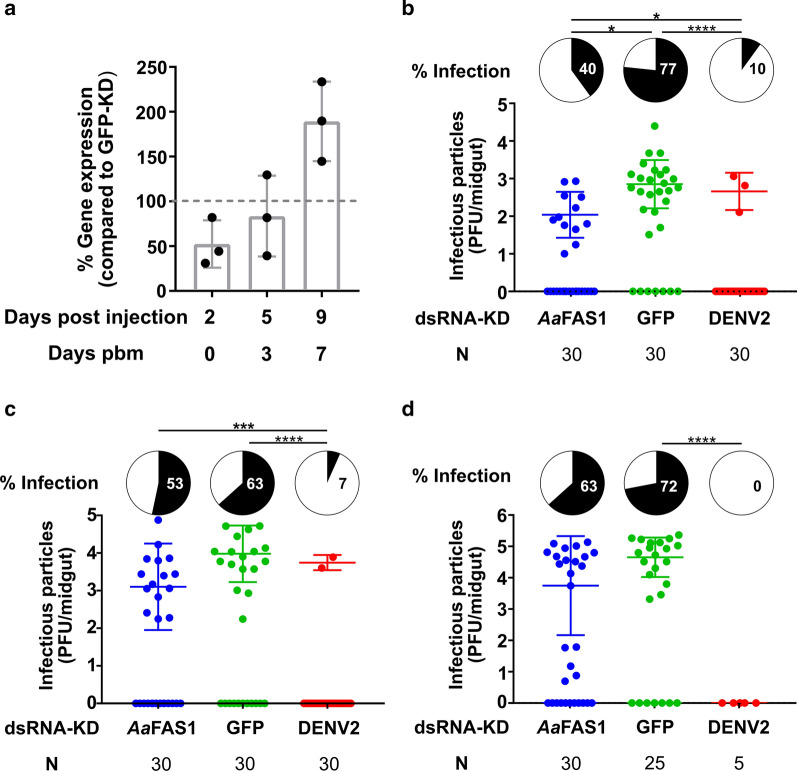
Fig. 8.
Transient KD of AaFAS1 expression by dsRNA temporarily reduced DENV2 infection in midguts. a Percent AaFAS1 expression in AaFAS1-KD compared to GFP-KD mosquitoes. Mosquitoes were IT injected with ~ 400 ng of dsRNA derived from AaFAS1 or GFP (negative KD control) and fed with a blood meal 2 days post IT injection. On days 2, 5 and 9 post IT injection (days 0, 3 and 7 pbm), 3 pools of 5 mosquitoes from both treatments were collected and analyzed for AaFAS1 expression. b–d Mosquitoes were IT injected with dsRNAs against AaFAS1, GFP and DENV2 (positive KD control) and infected with DENV2 via infectious blood meal at 2 days post injection. Plaque assay was performed on midguts dissected on (b) day 3 and (c) day 7 and (d) carcasses (whole body without midgut) collected on day 14 pbm. Pie charts (black) show percent infected mosquito tissue in each treatment. Pairwise χ2 tests with Holm’s correction for multiple comparisons were used to analyze differences in proportion of infected mosquitoes among groups. Dot plots report virus titer in mosquito tissues. Mean virus titer (infectious particles) was calculated for infected samples only. (i) and (ii) indicate the separation of DENV2 titers in the carcass (day 14) that were produced from the AaFAS1-KD mosquitoes. One-way ANOVA followed by Tukey’s multiple comparison tests were applied to test the differences in virus titer among samples but no significant differences in titers were found

