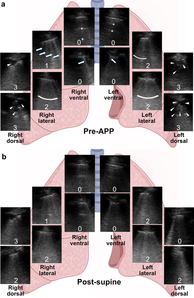
Fig. 4.
Typical changes in LUS score to APP in a patient with treatment success. a The patient’s LUS score at each lung zone before APP. Ventral zones had predominantly A-lines (asterisks) and ≤ 2 B-lines (arrows) (0 point) meaning normal aeration; lateral zones had ≥ 3 well-spaced B-lines (arrows) (1 point) and coalescent B-lines (curved bars) (2 points) suggesting moderate and severe aeration loss, respectively; and dorsal zones had irregular pleura, tissue-like pattern and subpleural consolidations (arrowheads) (3 points) suggesting complete loss of aeration. b The patient’s LUS score at each lung zone after returning to the supine position. Ventral, lateral, and upper right dorsal zones remained unchanged, while upper left dorsal zone improved from complete loss to normal aeration (3 to 0 points), and lower dorsal zones improved from complete to severe loss of aeration bilaterally (3 to 2 points). Total LUS score decreased from 19 to 14 in this patient whose first APP session lasted 5.5 h. The patient was in the supine position when LUS was performed. LUS, lung ultrasound; APP, awake prone positioning

