Fig. 3.
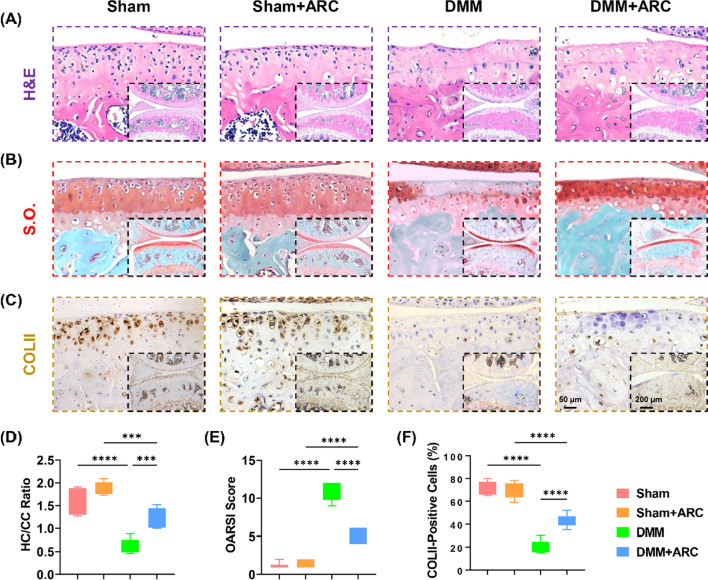
In vivo administration of arctiin mitigated DMM-induced articular cartilage abrasion in seven-week-old C57BL/6J male mice. A Representative images of articular cartilage stained by hematoxylin and eosin (H&E). B Representative images of articular cartilage stained by safranin O and fast green (S.O.). C Representative images of articular cartilage stained by collagen type II (COLII). D Quantification of the ratio of hyaline cartilage (HC) versus calcified cartilage (CC) (n = 6). E Quantification of the Osteoarthritis Research Society International (OARSI) score (n = 6). F Quantification of the percentage of COLII-positive cells in the articular cartilage (n = 6). Values represent the mean ± SD. Statistically significant differences are indicated by * where p < 0.05, ** where p < 0.01, *** where p < 0.001 and **** where p < 0.0001 between the indicated groups

