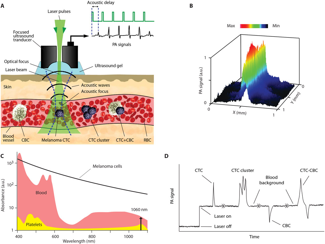
Fig. 1. The principle of CTC and CBC detection with the Cytophone platform using acoustic resolution PAFC.
(A) The schematic shows a focused ultrasound transducer with a central hole. Because of strong light scattering in tissue, the spatial resolution is determined by the acoustic focal volume. Laser-induced acoustic waves (referred to as PA signals) travel to the transducer with an acoustic time delay compared to laser pulse and PA background signals from the skin layer (23). (B) The lateral resolution (65 ± 6 μm) of the cylindrical transducer is shown as a PA signal distribution from black tape scanned with a focused laser beam with a diameter of 2 μm and a wavelength of 532 nm. a.u., arbitrary units. (C) The absorption spectra of RBCs (red), melanoma CTCs (black), and platelets (yellow) as the main components of white CBCs (7, 8, 23). (D) Left to right: PA trace with two positive PA peaks from a single CTC and a CTC cluster with three peaks above blood background and noise level, a negative PA peak from platelet-rich (white) CBC, and a peak with combined negative-positive contrast from CTC-CBC emboli (7, 23).

