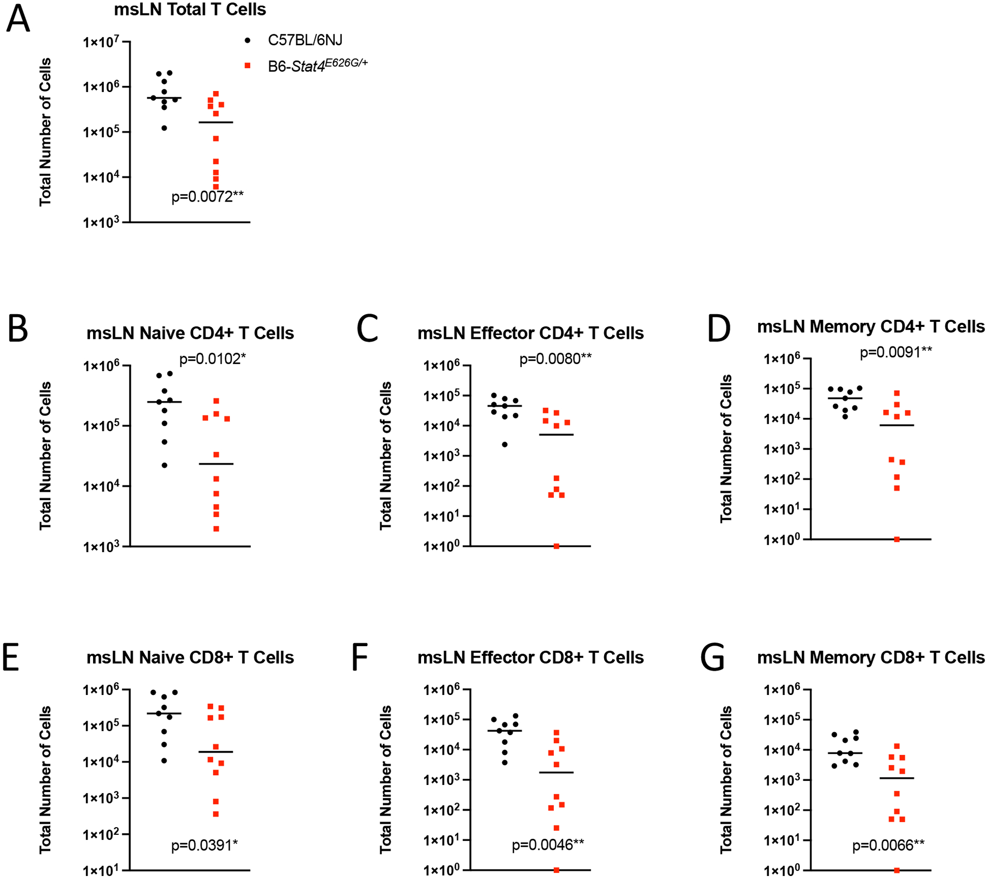
Fig. 6.

Stat4E626G/+ mice have decreased accumulation of total and activated T in the mediastinal lymph nodes 2 weeks after Cp1038 infection. C57BL/6NJ (black circles), B6-Stat4E626G/+ (red squares) mice were intranasally infected with Cp1038. Two weeks after infection mice were sacrificed (N=4–5/time point) and T cells were phenotyped in the mediastinal lymph nodes. (A) Total T Cells (CD3+ CD19−). (B,E) Naïve T cells (CD62L+ CD44var), (C,F) Effector T Cells (CD62L− CD44−), (D,G) Memory T Cells (CD62L− CD44+). Significance was determined by Mann-Whitney of log transformed data. Data is combined from 2 experiments of similar design.
