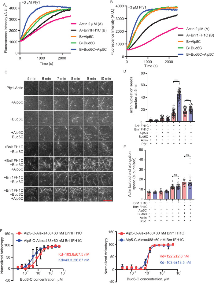
FIGURE 2:
Aip5 and Bud6 synergizes formin-mediated actin nucleation. (A, B) Pyrene actin polymerization with different combinations of proteins as indicated, 2 µM monomeric actin, 3 µM yeast profilin, 200 nM Aip5C, 40 nM Bud6C, 20 nM Bni1FH1C, and 20 nM Bnr1FH1C. (C) The representative TIRF images of actin nucleation seeds formed from 5 to 10 min using the indicated combination of proteins. The actin filament was assembled by mixing 1 µM actin (10% Oregon green 488-labeled actin and 0.5% biotin-actin) with 3 µM yeast profilin, 10 nM Bni1FH1C, 2 nM Bnr1FH1C, 5 nM Bud6C, or 20 nM Aip5C. The scale bar represents 10 µm. (D) Quantification of actin nucleation seeds number at 5 min with the indicated combinations of proteins (n = 80, 60, 98, 94, 114, 99, 100, 100 for each sample from ROI = 894 µm2). (E) Quantification of actin filament barbed end elongation speed of indicated protein combinations as shown in A (n = 28, 21, 24, 23, 27, 43, 19, 37 for each sample). (F) Fluorescence anisotropy binding measurements of 60 nM Alexa 488-labeled Aip5C were preincubated with different concentrations of Bni1FH1C and Bni1FH1C, respectively, before being titrated by a serial concentration of Bud6C. P values was determined by the one-way ANOVA, ns = not significant, ****p < 0.0001. Error bar, SD.

