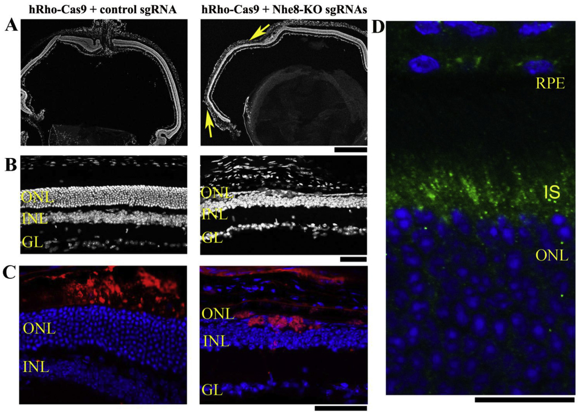
Fig. 4. Photoreceptor specific knockdown of NHE8 by AAV-hRho-NmCas9 and AAV-Nhe8-KO sgRNAs caused retinal degeneration in adult wild-type mice.

(A–C) The left panels are retinal sections of a control eye injected with a mixture of AAV-hRho-Cas9 and AAV-control sgRNA; the right panels are retinal sections from the right eye of the same mouse injected with AAV-hRho-Cas9, AAV-Nhe8-KO sgRNAs-P1/P2 and AAV-Nhe8-KO sgRNAs-E2/E5/E6. (A) A comparison of low magnification images of DAPI-stained retinal frozen sections between control and knockdown eyes. Two arrows (yellow) in the right image indicate areas with loss of photoreceptor cells. (B) DAPI-stained frozen retinal sections showed control on the left and the knockdown on the right with only one single layer of photoreceptor cells. (C) Images of retinal frozen sections co-stained with anti-HA for Cas9 protein (red) and DAPI (blue). HA tag was constructed in-frame into the C-terminus of Cas9 in the vector, thus the AAV-Cas9 expression can be traced using an antibody recognizing HA tag. ONL: outer nuclear layer; INL: inner nuclear layer; GL: ganglion cell layer. Scale bars: upper panels, 500 μm; middle and lower panels, 50 μm. (D) A retinal section of 3 weeks-old wild-type mouse stained with NHE8 antibody (green) and Dapi (blue). Endogenous NHE8 staining signals were observed in the retinal pigment epithelial cells (RPE) and inner segments (IS) of photoreceptor cells. Scale bar: 20 μm.
