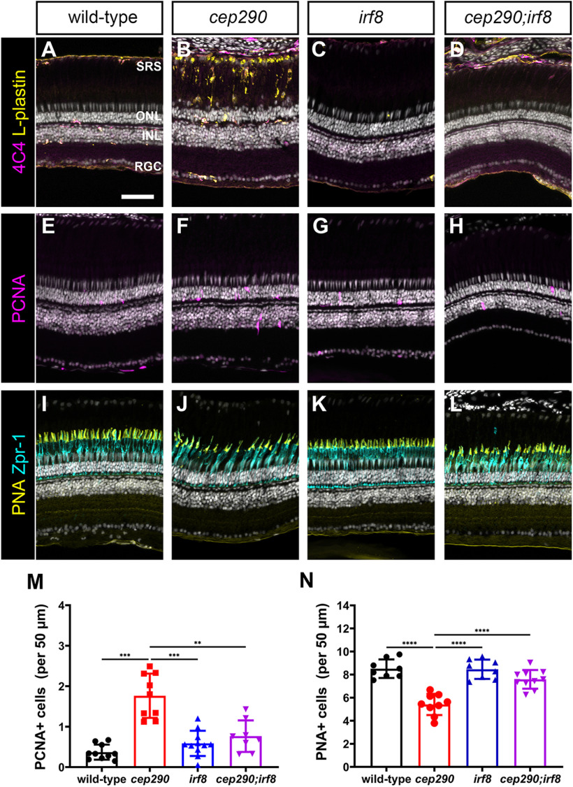
Figure 11.
Mutation of irf8 reduces the number of activated microglia/macrophage and proliferating cells and promotes cone survival in cep290 mutants. A–D, Immunohistochemistry of the dorsal retina with markers for microglia/macrophage (4C4, magenta) and leukocytes (L-plastin, yellow) in 6 mpf wild-type, cep290 mutants, irf8 mutants, and cep290;irf8 mutants. E–H, Immunohistochemistry of the dorsal retina at 3 d postinjection with PCNA (magenta) to mark proliferating cells in 6 mpf wild-type, cep290 mutants, irf8 mutants, and cep290;irf8 mutants. I–L, Immunohistochemistry of the dorsal retina with markers for cone inner segments (Zpr-1, cyan) and cone outer segments (PNA, yellow) in 6 mpf wild-type, cep290 mutants, irf8 mutants, and cep290;irf8 mutants. M, N, Quantification of PCNA+ cells in the ONL and PNA+ outer segments in wild-type (filled black circles) and cep290 mutants (filled red circle), irf8 mutants (filled blue triangles), and cep290;irf8 mutants (filled purple triangles). Each data point represents counts from the dorsal retina of one eye. For the graph in M, the significance in differences was determined using Welch ANOVA test with Dunnett's T3 multiple comparisons test (**p < 0.01, ***p < 0.001). For the graph in N, the significance in differences was determined by one-way ANOVA with Tukey's multiple comparisons test (***p < 0.001, ****p < 0.0001). SRS = subretinal space; ONL = outer nuclear layer; INL = inner nuclear layer, RGC = retinal ganglion cell layer. Scale bars: 400 µm (A, B) and 50 µm (D–O).

