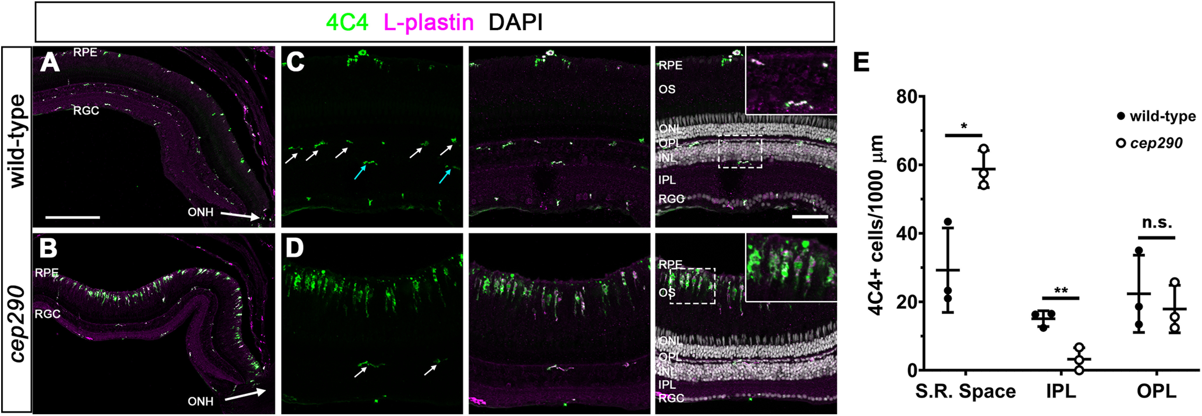
Figure 2.

Immune cells accumulate in the subretinal space of cep290 mutants. A, B, Immunohistochemistry with the monoclonal antibody 4C4 (green) and anti-L-plastin (magenta) label microglia/macrophage in the dorsal retina of 6 mpf wild-type and cep290 mutants. The ONH is located at the bottom right corner of each image. C, D, Higher magnification images show the accumulation of activated microglia/macrophage in the subretinal space of cep290 mutants. Ramified microglia/macrophage (4C4+/L-plastin+) were seen in the OPL (white arrows) of both wild-type and cep290 mutants but only in the IPL of wild-type retinas (cyan arrows). Insets, Higher magnification of boxed regions illustrates the ramified morphology of quiescent microglia/macrophage in the plexiform layers of wild-type retinas and the elongated and amoeboid shape of microglia/macrophage in the subretinal space of cep290 mutants. E, Quantification of 4C4+ cells in different regions of the retina in 6 mpf fish (n = 3 per genotype). Data are plotted as mean ± SD and p-values were generated by Welch's t tests. *p < 0.05, ****p<0.01; n.s. = not significant. RPE = retinal pigment epithelium, RGC = retinal ganglion cell layer, ONH = optic nerve head, OS = outer segments, ONL = outer nuclear layer, INL = inner nuclear layer, S.R. space = subretinal space, IPL = inner plexiform layer, OPL = outer plexiform layer. Scale bars: 200 µm (A, B) and 50 µm (C, D).
