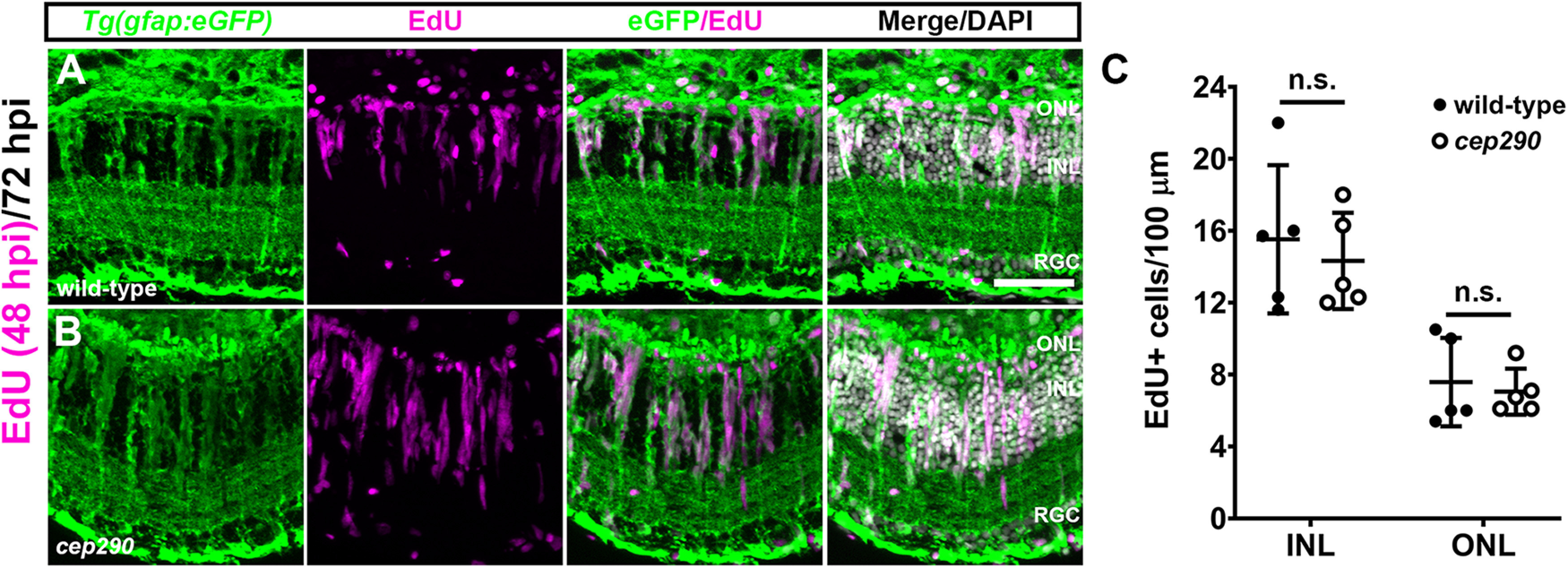
Figure 6.

MG in cep290 mutants respond to acute light damage. Retinal cryosections of 6 mpf wild-type (A) or cep290 mutants (B) carrying the Tg(gfap:eGFP)mi2002 transgenic reporter line were immunolabeled with anti-GFP (green) antibodies to visualize MG and processed for EdU labeling (magenta) to identify MG-derived progenitor cells at 72 hpi. C, The number of EdU+ cells was not statistically different (n.s.) between wild-type and cep290 mutants (INL: p > 0.99; ONL: p > 0.66; Mann–Whitney tests). Data are plotted as mean ± SD ONL = outer nuclear layer; INL = inner nuclear layer, RGC = retinal ganglion cell layer. Scale bar: 50 µm.
