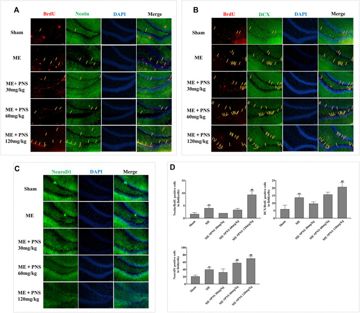
FIGURE 5.
PNS administration stimulated post-ischemic hippocampal neurogenesis in ME rats. Representative images of ipsilateral hemisphere sctions under a fluorescence microscope at a magnification of ×200 (scale bar =100 μm) were shown as (A) BrdU (red, a marker of proliferating cells) and Nestin (green, a marker of NSC/NPC); (B) BrdU (red) and DCX (green, a marker of migrating neuroblasts); (C) NeuroD1 (green, a marker of differentiation factor). (D) The numbers of double-positive of BrdU/Nestin, BrdU/DCX, and single-positive NeuroD1 were analyzed and data were presented as means ± standard deviation (n = 3 animals per group). *p < 0.05, **p < 0.01 versus Sham group; # p < 0.05, ## p < 0.01 versus ME group.

