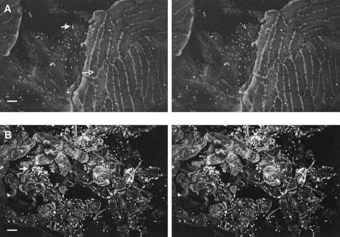
FIG. 8.
CSLM stereo images showing attachment of E. coli O157:H7 to the ventral cavity. (A) More attached cells (closed arrow) were observed in crevices (38-μm depth) than on the smooth regions of cartilaginous pericarp (open arrow). (B) Irregular tissue (42-μm depth) on the ventral cavity harboring many cells (closed arrow). Bar, 10 μm.

